Skip to main content
Fish Pathogens
Menu
Search
Search
Main navigation
Image Galleries
Pathogens
Pathogens sub-navigation
10 Common Pathogens
What's That Spot?
Ask an Expert
FAQ
Search
Search
Search
User account menu
Log in
Primary tabs
Common Pathogens
All Pathogens
Hosts
Field Sites
Videos
Contributed
All Pathogens
These are all of the pathogen images in our database. Click a pathogen name to jump to the pathogen info page. Click an image to enlarge it, then use the left and right arrows to browse.
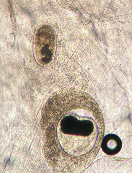
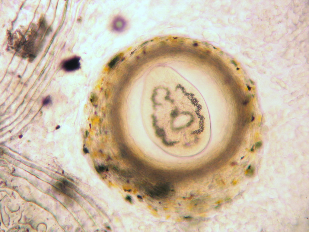
Posthodiplo…
EIBS
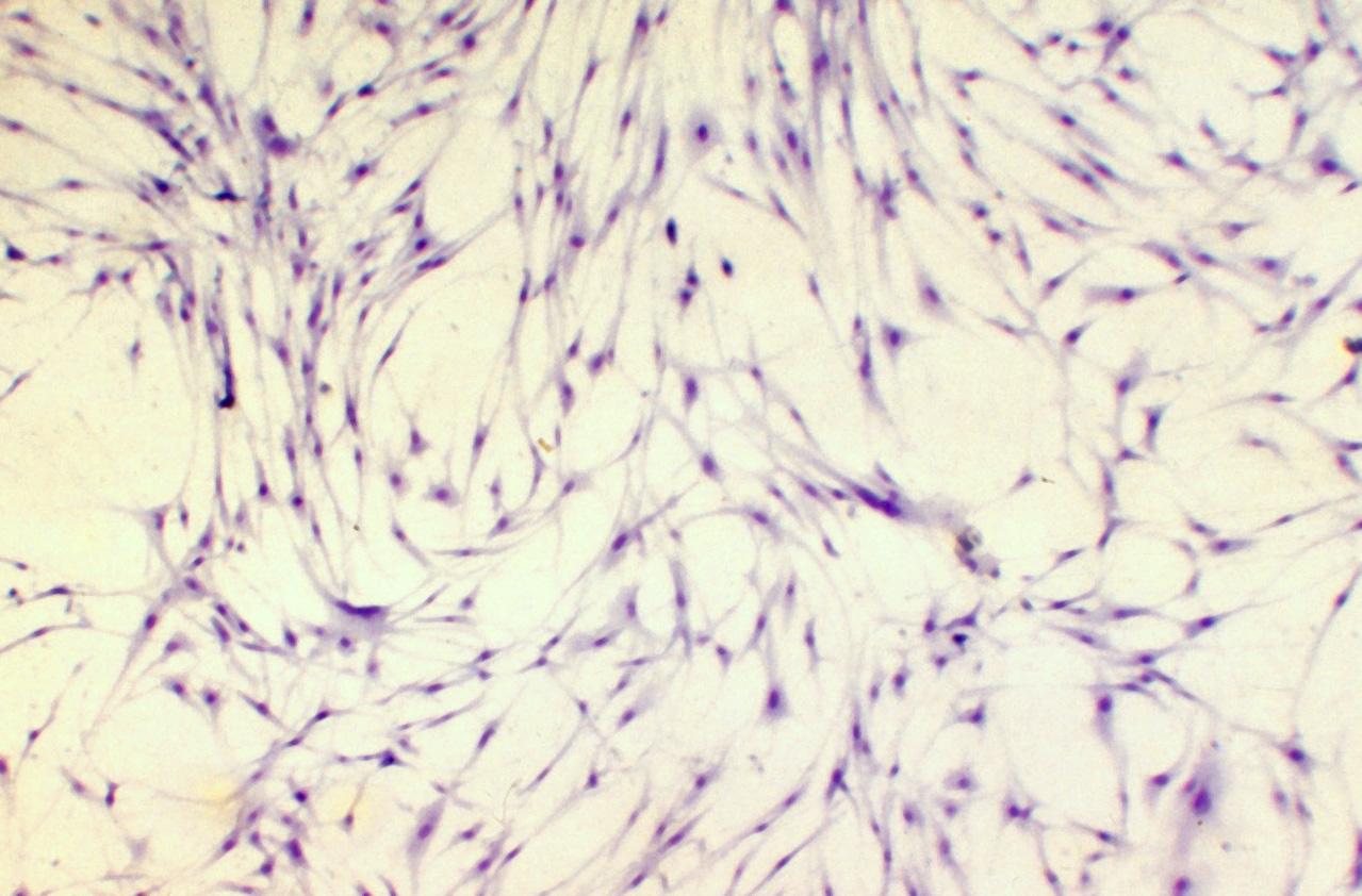
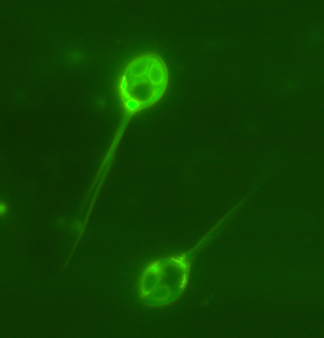
Chilodonella
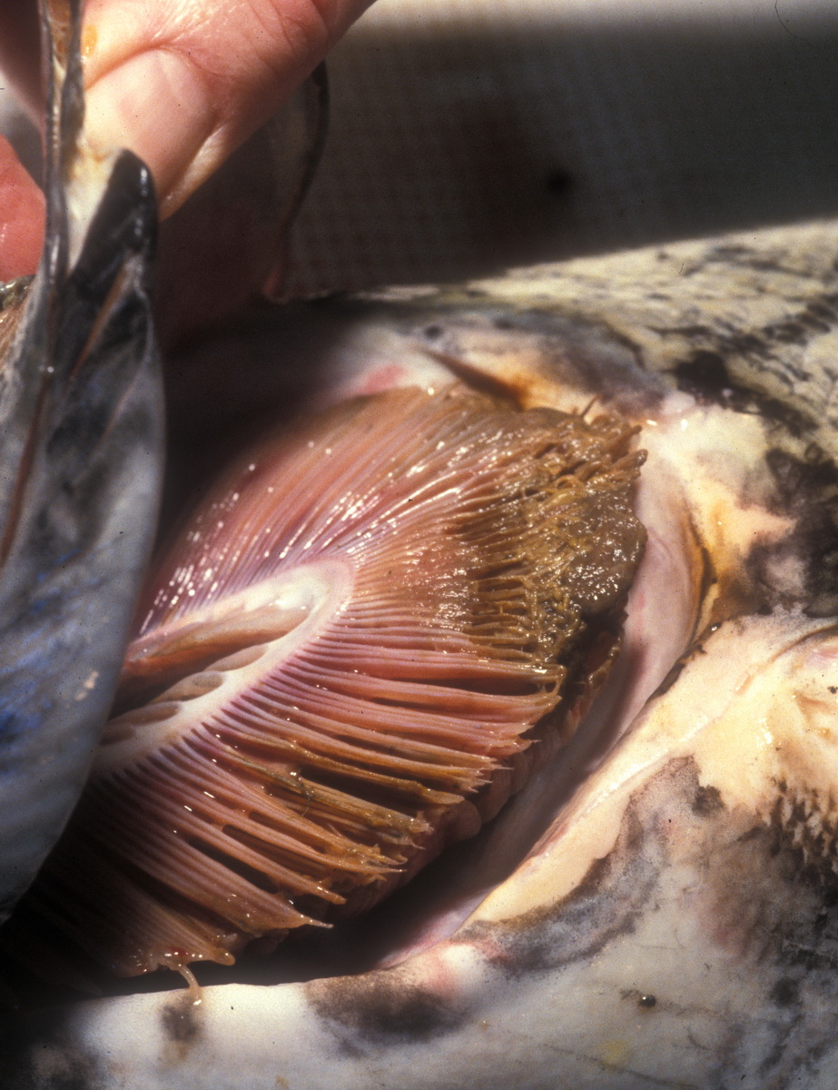
Aeromonas salmonic…
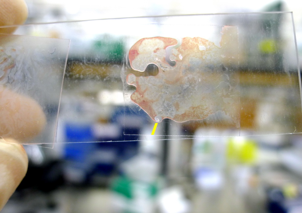
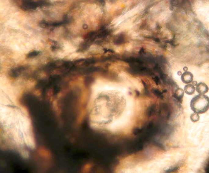
Dermocystidium
Epistylis
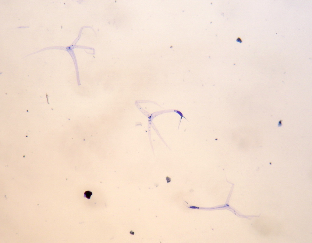
Copepods
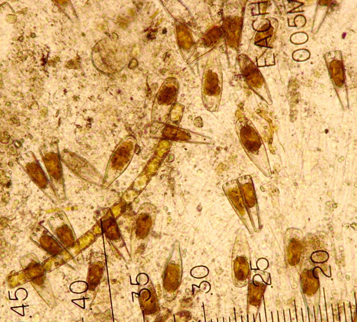
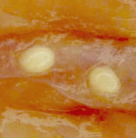
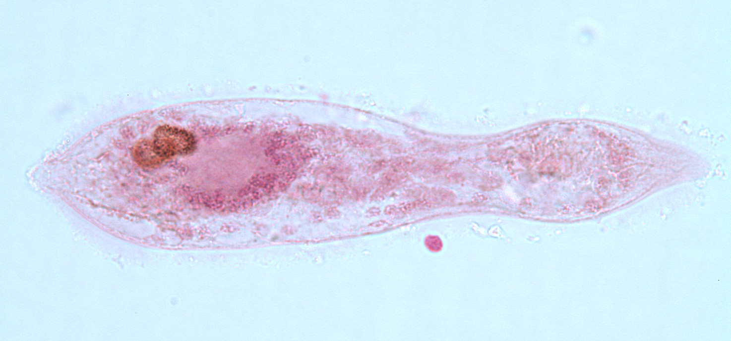
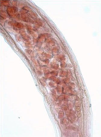
Trematodes (fluk…
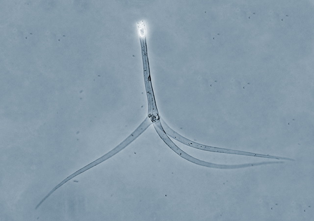
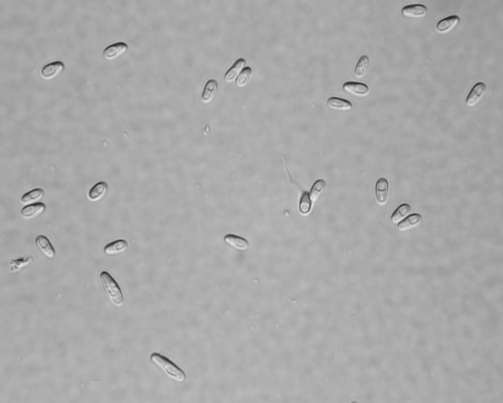
…
Henneguya salminic…
Vibrio
Myxobolus squamalis…
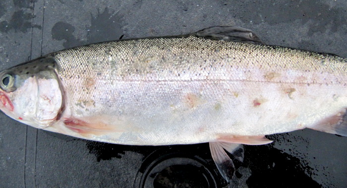
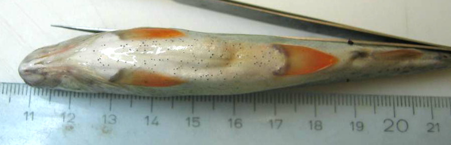
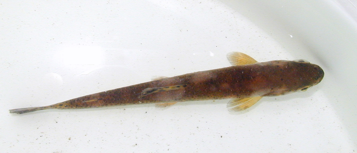
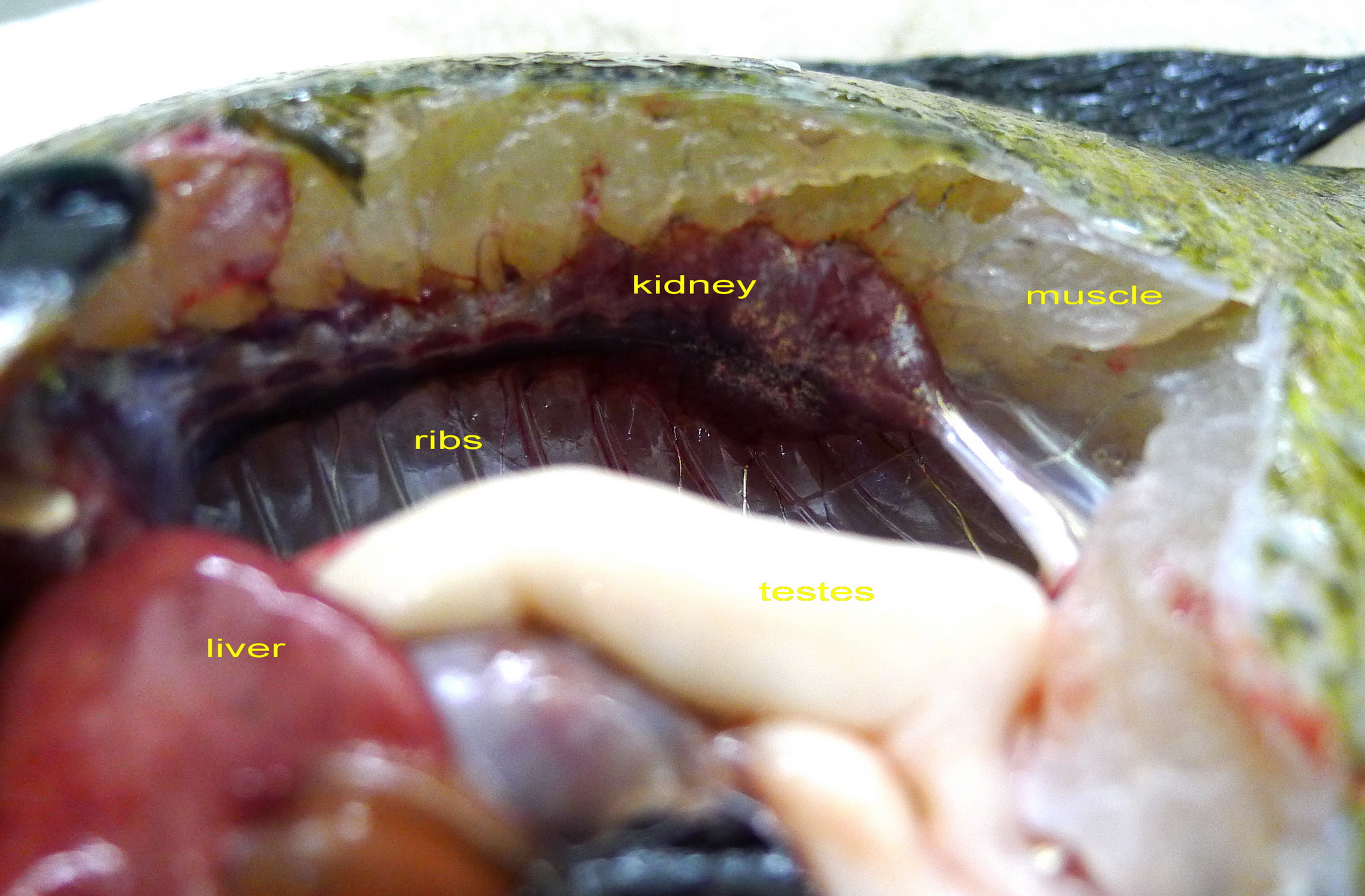
White Sturgeon…
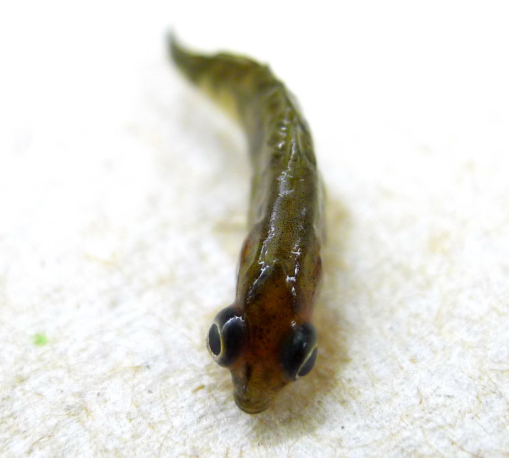
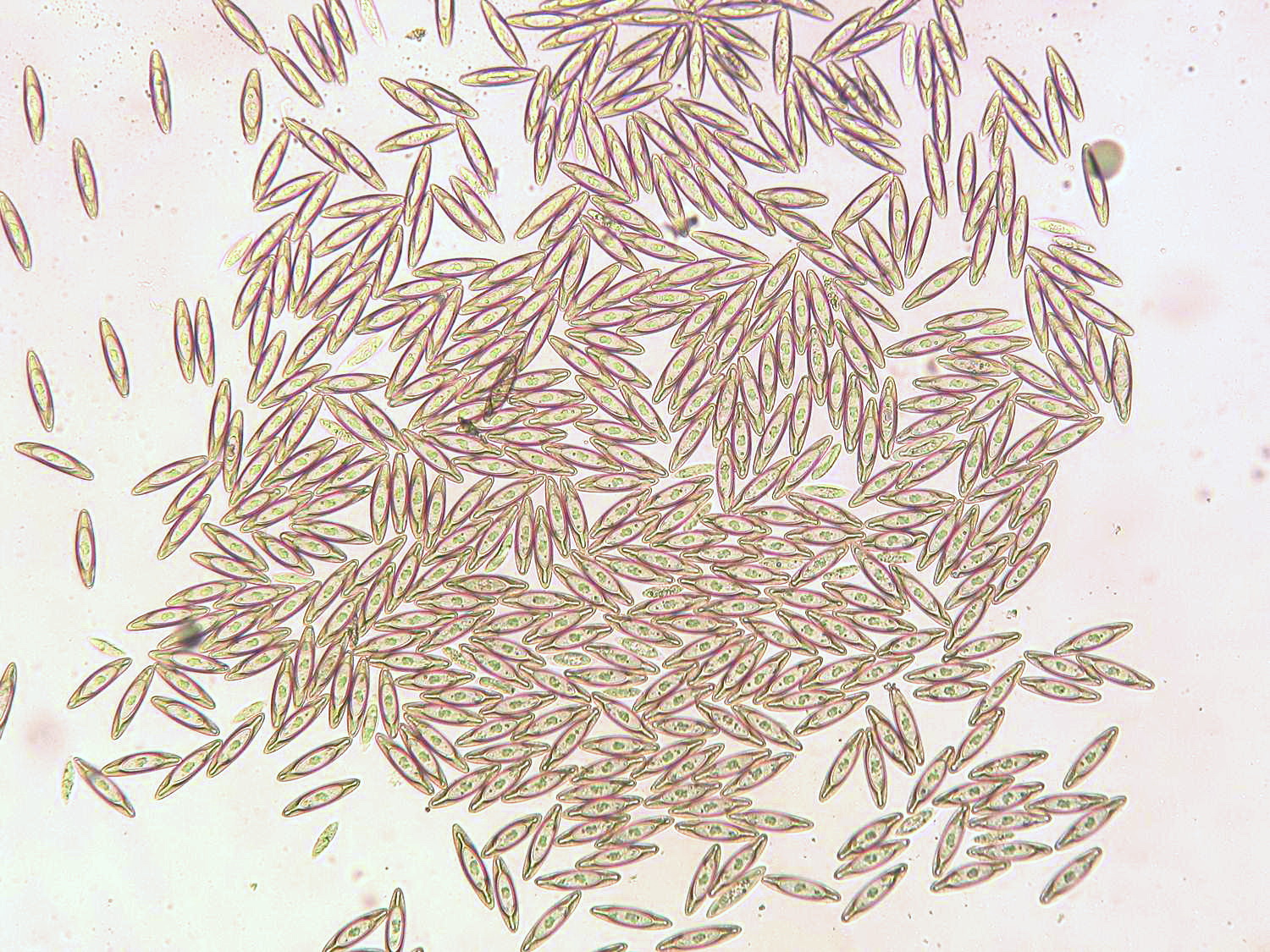
Nanophyetus
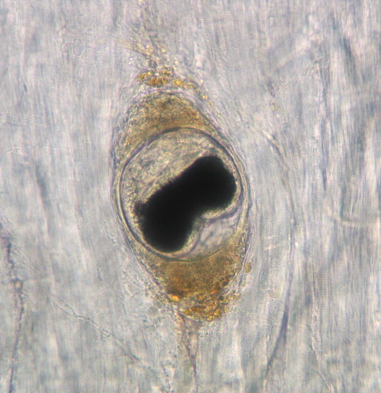
…
Trematodes (fluk…
Mycobacterium mari…
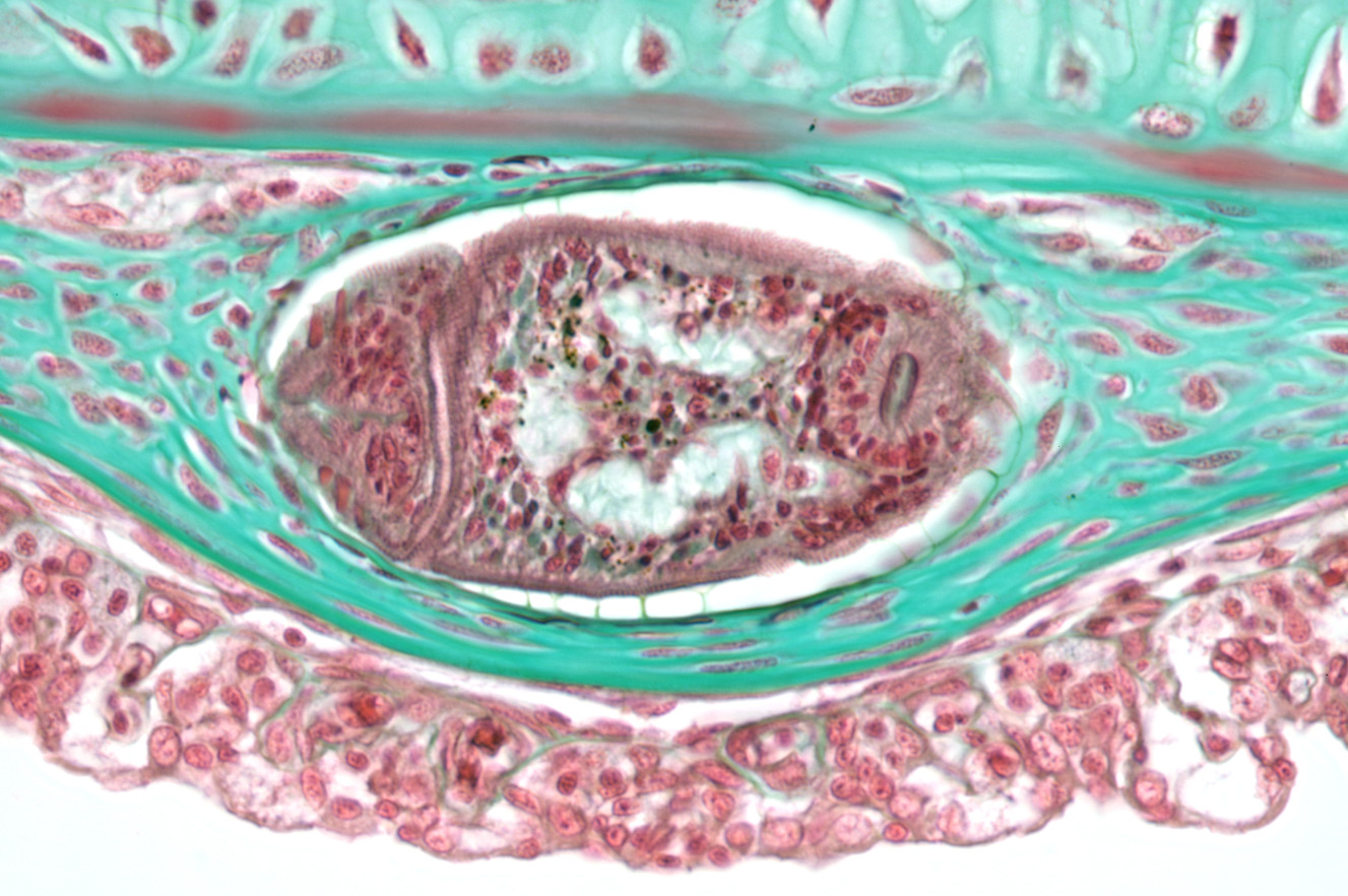
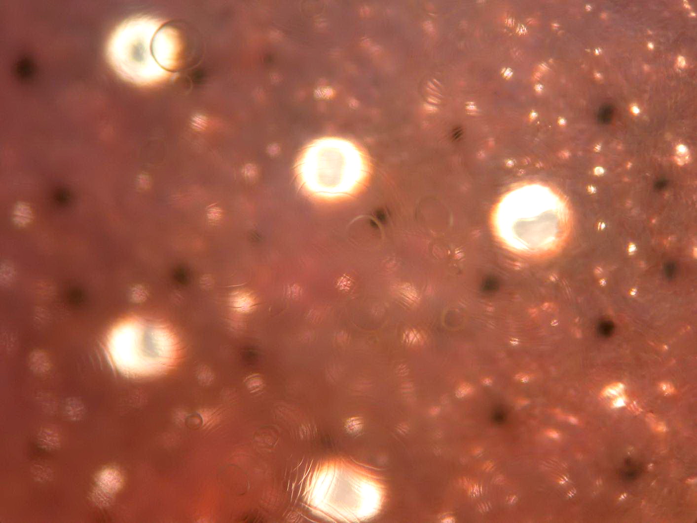
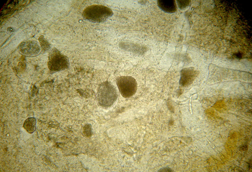
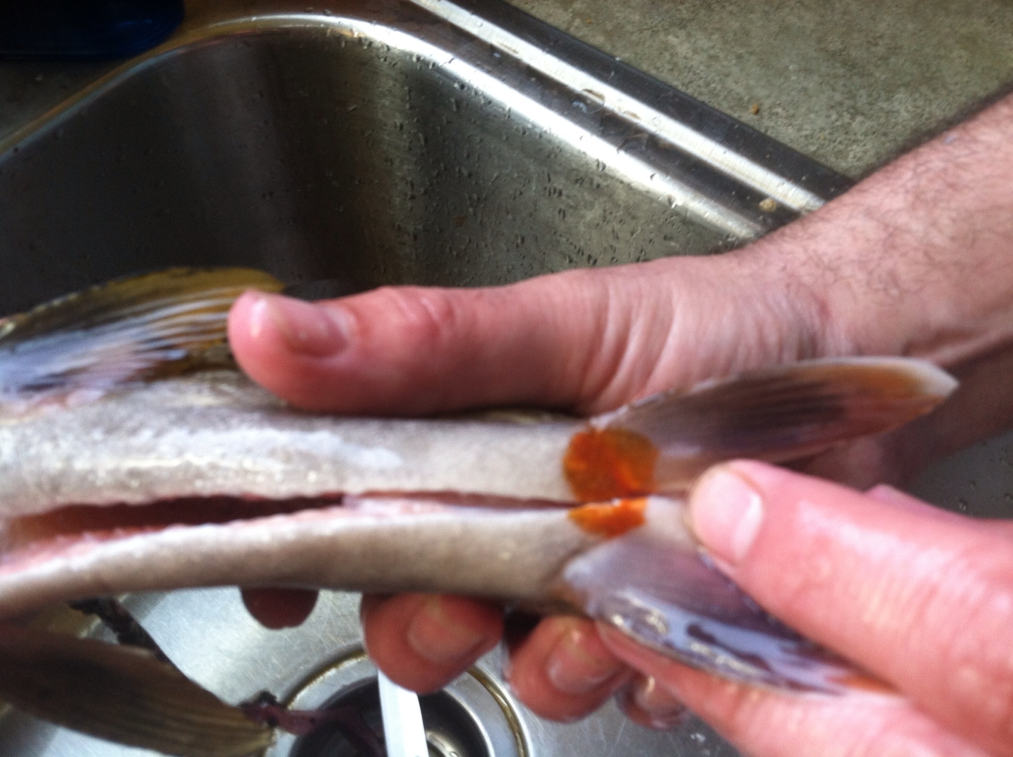
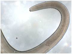
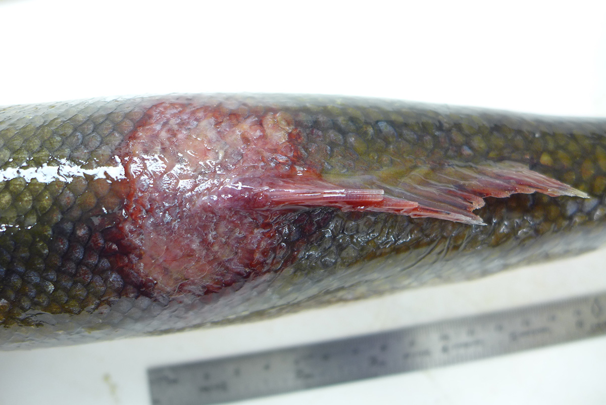
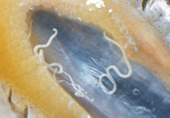
Philometroides
Henneguya salminic…
Sanguinicola
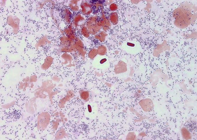
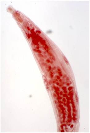
Trypanosomes
Nanophyetus
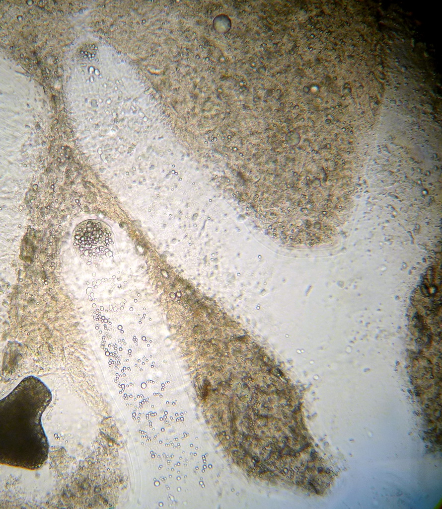
Ichthyophonus
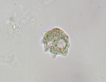
Flav…
non-pa…
Trematodes (fluk…
Neoechinorhynchus s…
Trematodes (fluk…
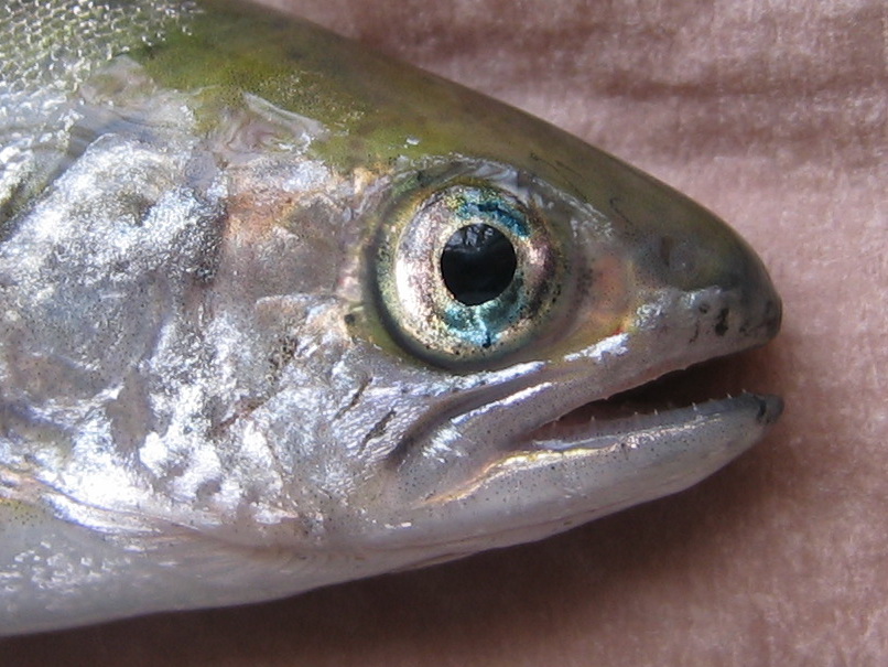
Anisakis
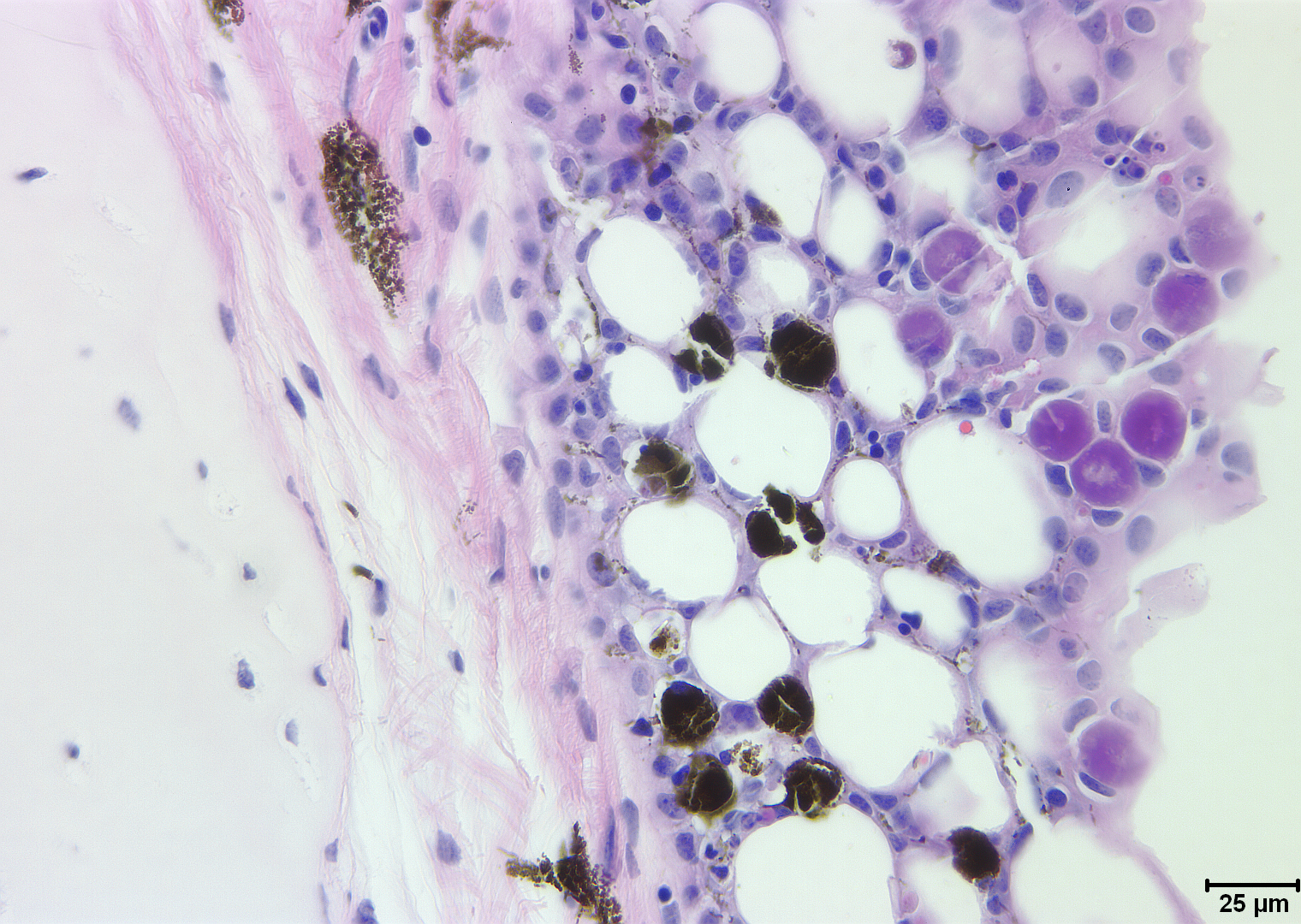
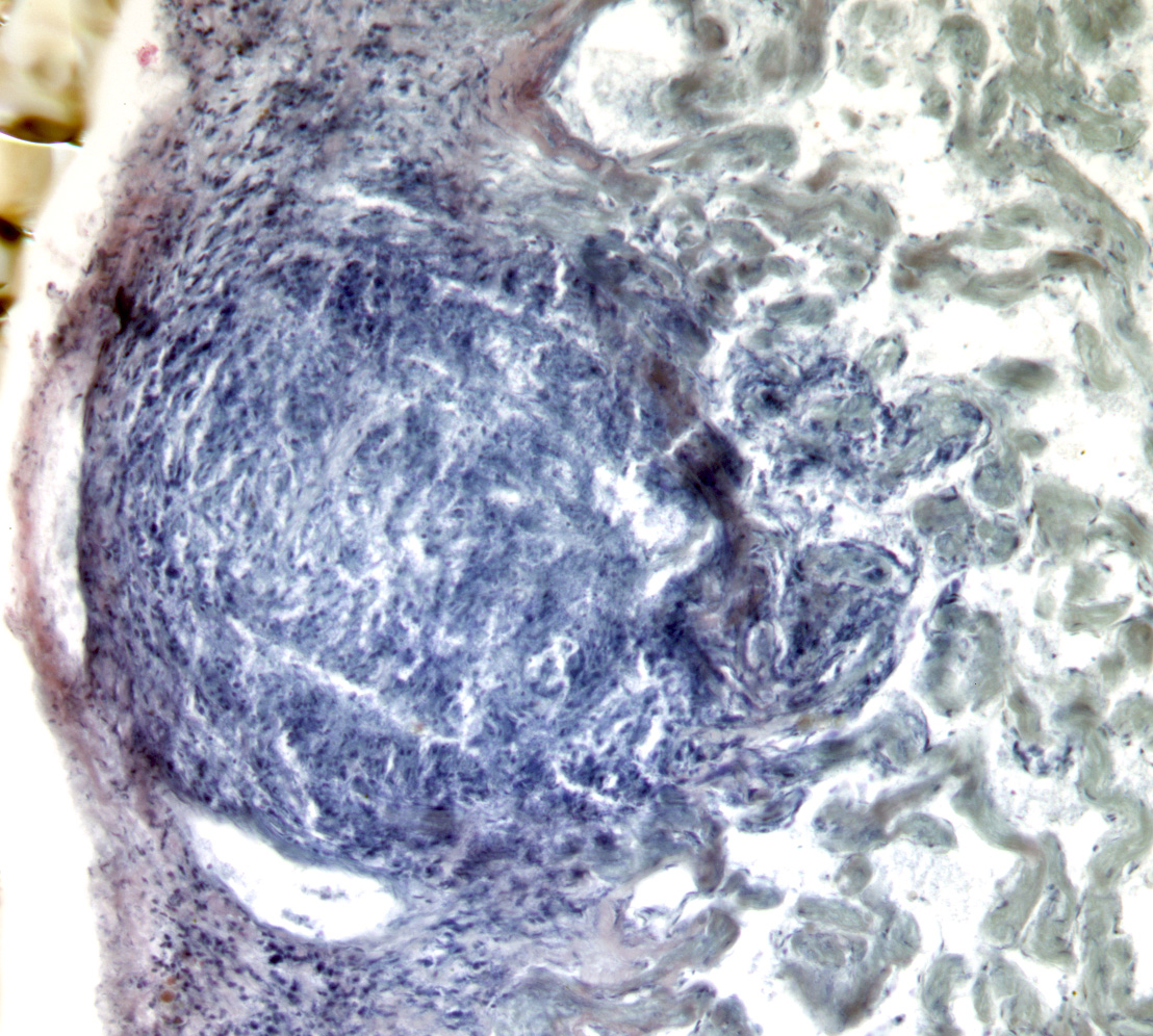
Myxobolus cerebrali…
Neascus
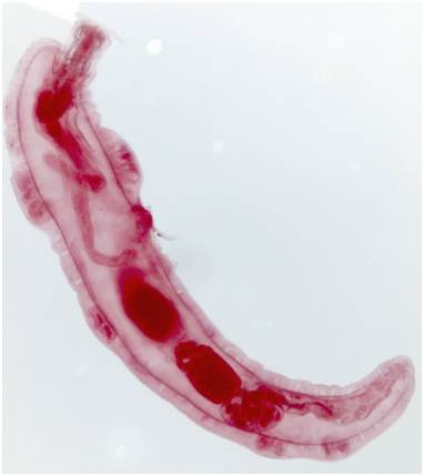
Aca…
Fungi and fun…
Trematodes (fluk…
Apiosoma
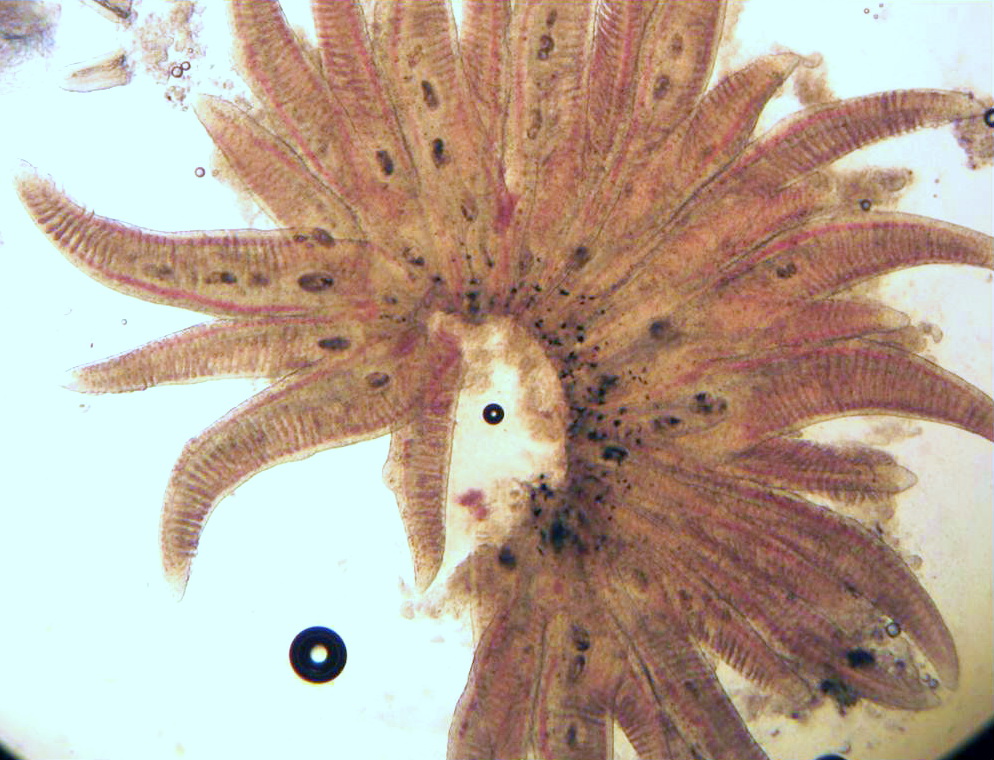
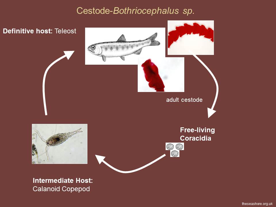
Diphyllob…
Nanophyetus
…
Neoechinorhynchus s…
Echinochasmus
Trematodes (fluk…
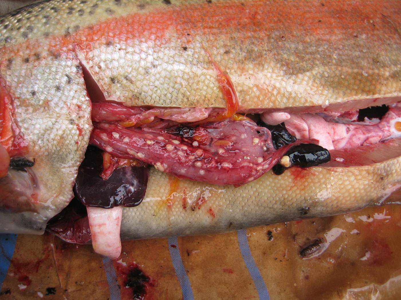
Philonema
Bacteria
Trematodes (fluk…
Neoechinorhynchus s…
Ichthyophonus
Cystidicola
Pagination
Previous page
2
Next page
RSS feed