 | DSC00263_amandi.jpg | Personel, Fact Sheet Images | |
 | P1050695_sampling_CogswellCk.jpg | Field Localities (-> and enter Locality below), Personel, Fact Sheet Images | |
 | DSC00494_bjork.jpg | Personel, Fact Sheet Images | |
 | DSC00495_arsan.jpg | Personel, Fact Sheet Images | |
 | Banner_P1050452.jpg | Personel, Equipment: laboratory / tanks / methods..., Fact Sheet Images | rainbow trout |
 | olsenb.gif | Personel, Fact Sheet Images | |
 | Banner_P1050453.jpg | Personel, Equipment: laboratory / tanks / methods..., Fact Sheet Images | rainbow trout |
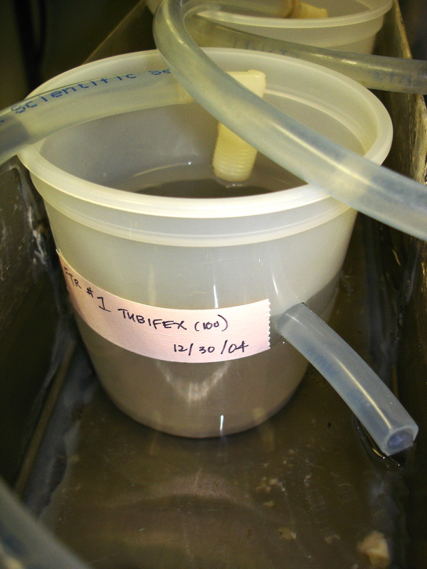
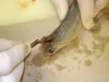
 | SLH_DSC00305.jpg | Fact Sheet Images, Hosts and other organisms, Equipment: laboratory / tanks / methods... | Tubifex |
 | SLH_163_6368.jpg | Fact Sheet Images, Equipment: laboratory / tanks / methods... | |
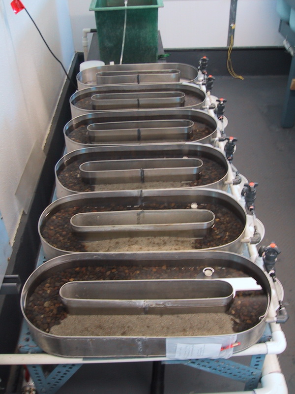
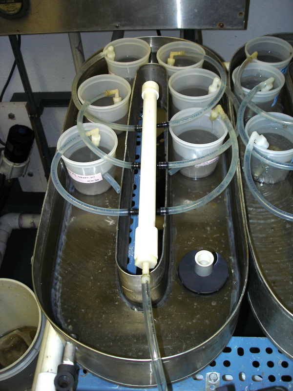
 | SLH_DSC01938.jpg | Equipment: laboratory / tanks / methods... | rainbow trout |
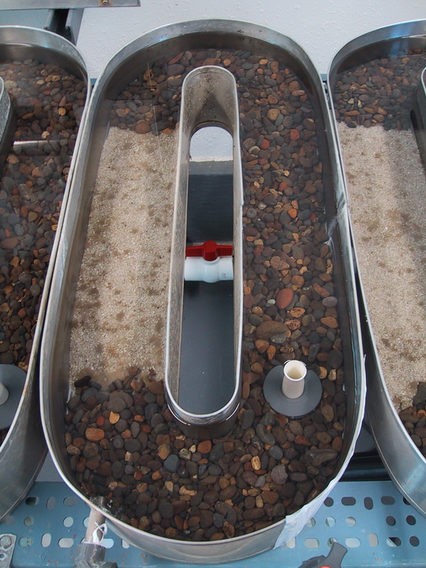
 | SLH_172_7220.jpg | Fact Sheet Images, Equipment: laboratory / tanks / methods... | |
 | SLH_DSC01940.jpg | Equipment: laboratory / tanks / methods... | rainbow trout |
 | SLH_DSC00522.jpg | Fact Sheet Images, Equipment: laboratory / tanks / methods... | |
 | SLH_DSC01941.jpg | Equipment: laboratory / tanks / methods... | rainbow trout |
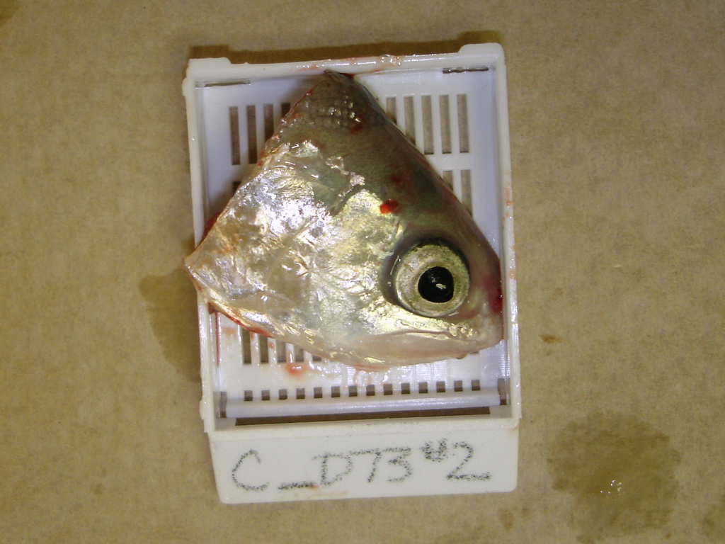
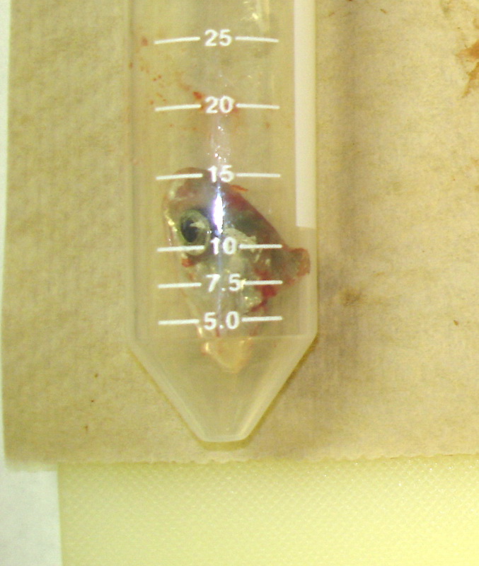
 | SLH_100_0018_r1.jpg | Hosts and other organisms, Equipment: laboratory / tanks / methods..., Fact Sheet Images | rainbow trout |
 | SLH_DSC00059.jpg | Fact Sheet Images, Equipment: laboratory / tanks / methods... | |
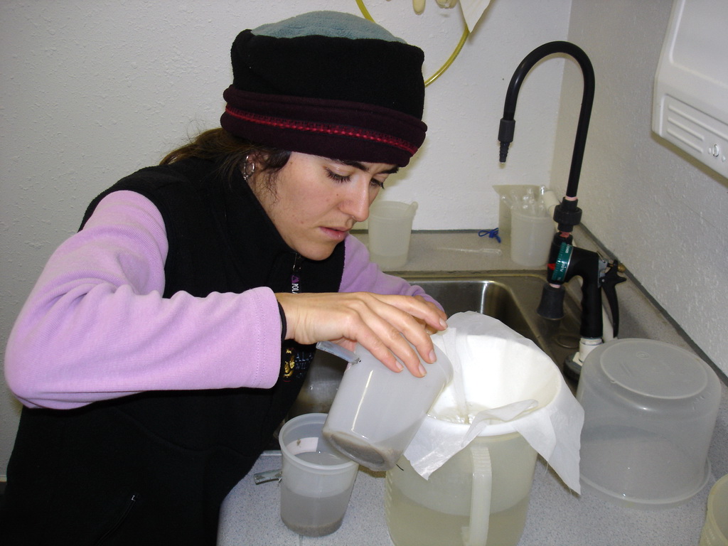
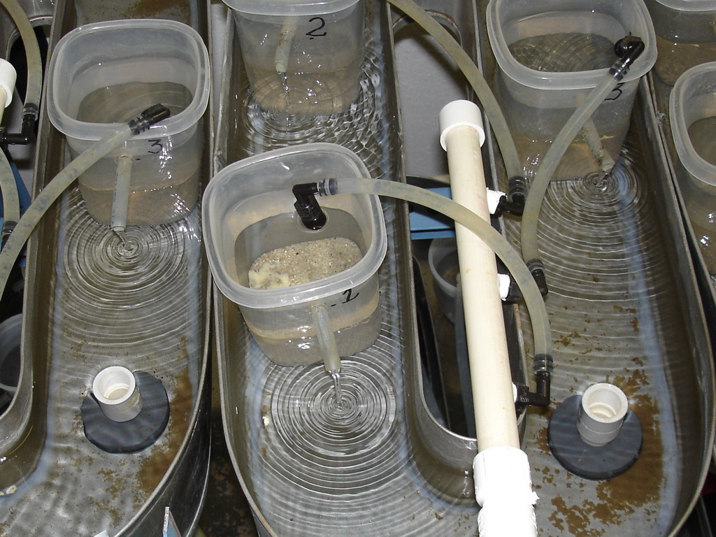
 | SLH_DSC00628.jpg | Equipment: laboratory / tanks / methods... | Tubifex |
 | SLH_DSC00339.jpg | Equipment: laboratory / tanks / methods... | Tubifex |
 | SLH_DSC00060.jpg | Fact Sheet Images, Equipment: laboratory / tanks / methods... | |
 | SLH_DSC00340.jpg | Equipment: laboratory / tanks / methods... | Tubifex |
 | SLH_DSC00331.jpg | Equipment: laboratory / tanks / methods... | Tubifex |
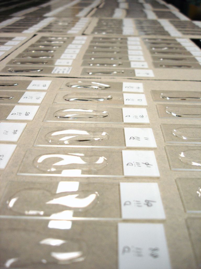
 | SA_DSC03909.jpg | Equipment: laboratory / tanks / methods..., Fact Sheet Images | Tubifex |
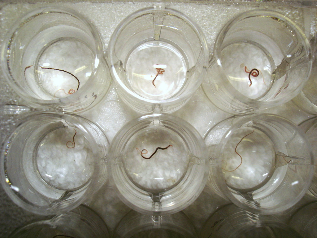
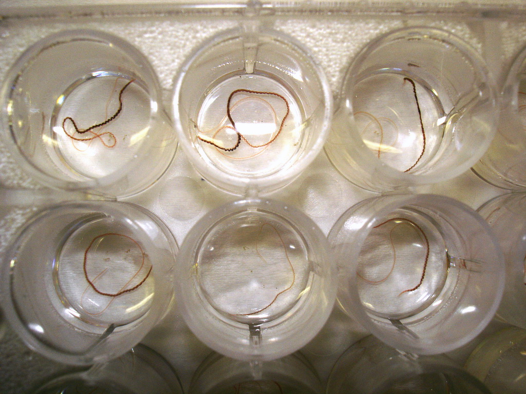
 | SLH_DSC00266.jpg | Fact Sheet Images, Hosts and other organisms, Equipment: laboratory / tanks / methods... | Tubifex |
 | SLH_DSC00312.jpg | Hosts and other organisms, Equipment: laboratory / tanks / methods... | Tubifex |
 | SLH_DSC00191.jpg | Fact Sheet Images, Equipment: laboratory / tanks / methods... | |
| | Banner_P1050452.jpg | Personel, Equipment: laboratory / tanks / methods..., Fact Sheet Images | rainbow trout |
 | SLH_DSC00281.jpg | Fact Sheet Images, Hosts and other organisms, Equipment: laboratory / tanks / methods... | Tubifex |
 | SLH_DSC00259.jpg | Fact Sheet Images, Equipment: laboratory / tanks / methods... | |
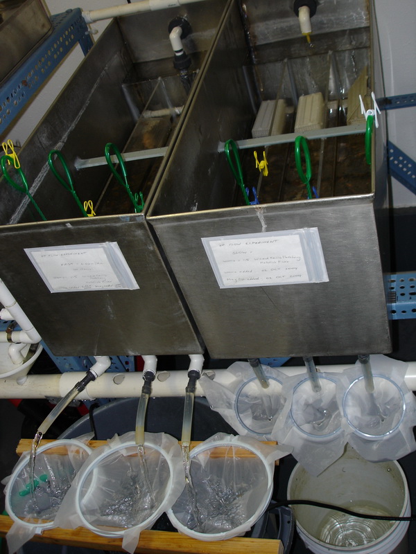
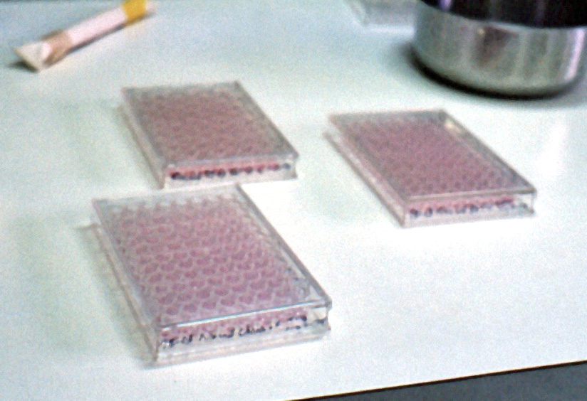
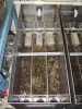
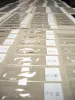
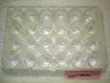
 | JJ0002_SAed_cell_culture_plates_for_virus.jpg | Equipment: laboratory / tanks / methods... | |
 | SLH_163_6367.jpg | Fact Sheet Images, Equipment: laboratory / tanks / methods... | |