 | CB_IMG_5021.jpg | All Pathogens, Fact Sheet Images | |
 | CB_HeavyIch2.jpg | All Pathogens, Fact Sheet Images | |
 | CB_IMG_0501.jpg | All Pathogens, Fact Sheet Images | |
 | CB_TumorCoRC.jpg | All Pathogens, Fact Sheet Images | coho salmon |
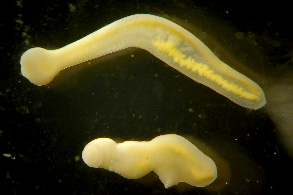
 | SLH_DSC00212.jpg | All Pathogens | Tubifex |
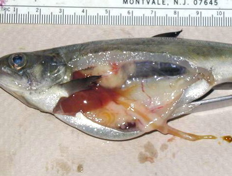
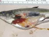
 | P1000375ed.jpg | All Pathogens, Fact Sheet Images | smallmouth bass |
 | CB_BKD_02_ChS.jpg | All Pathogens, Fact Sheet Images | Spring Chinook salmon |
 | IHNV_kaufman1.jpg | All Pathogens, Fact Sheet Images | |
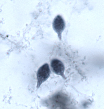
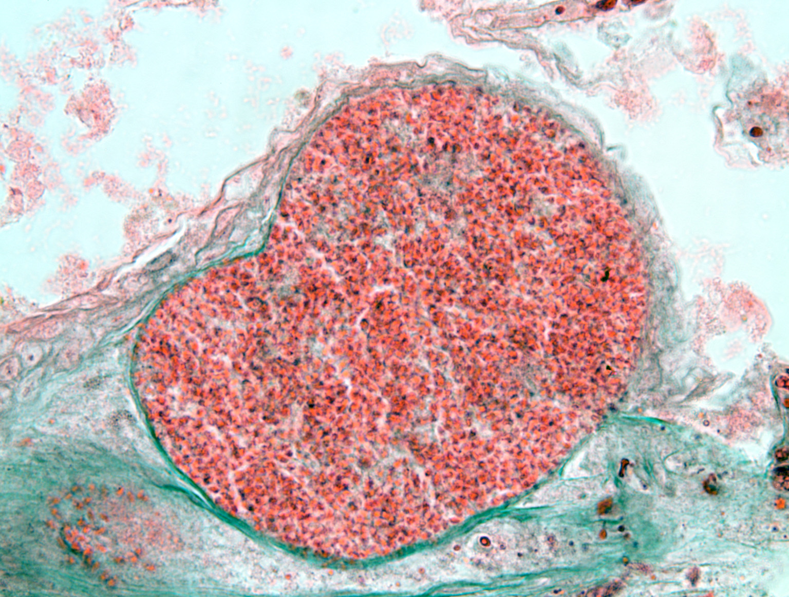
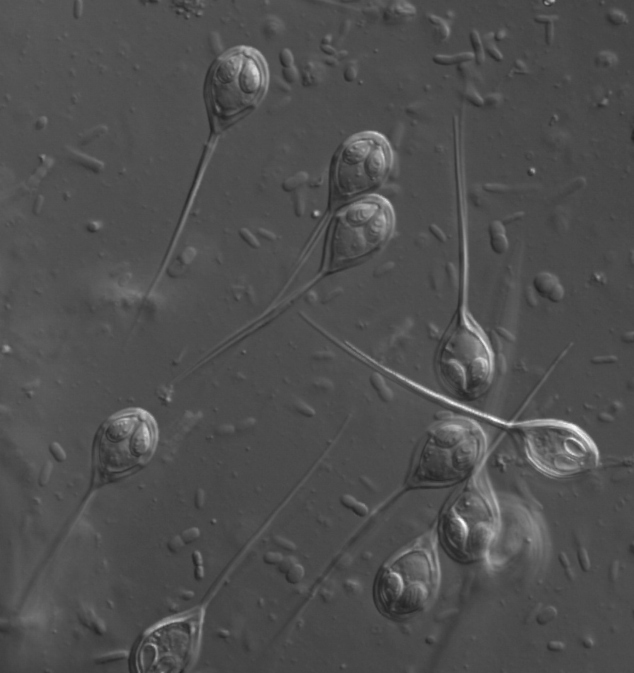
 | BO_Dermocystidium_x63_25.jpg | All Pathogens, Fact Sheet Images | |
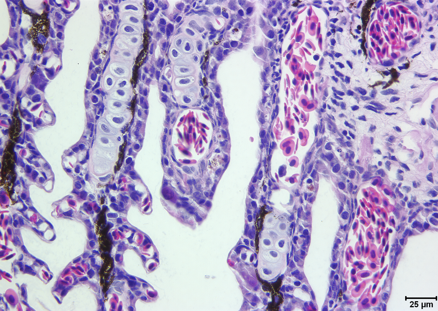
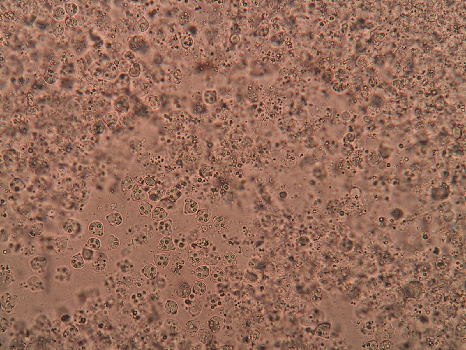
 | BO_Hexamita_x63_36.jpg | All Pathogens, Fact Sheet Images | |
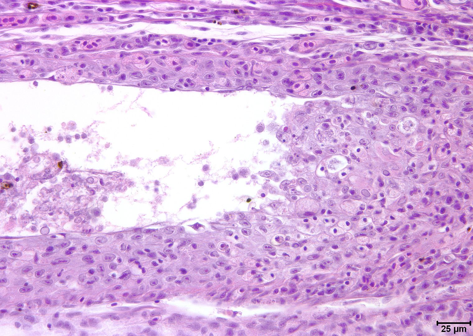
 | Slide-#-100-Gills-Koi-Herpes-Virus-H&E-400X.jpg | All Pathogens | common carp |
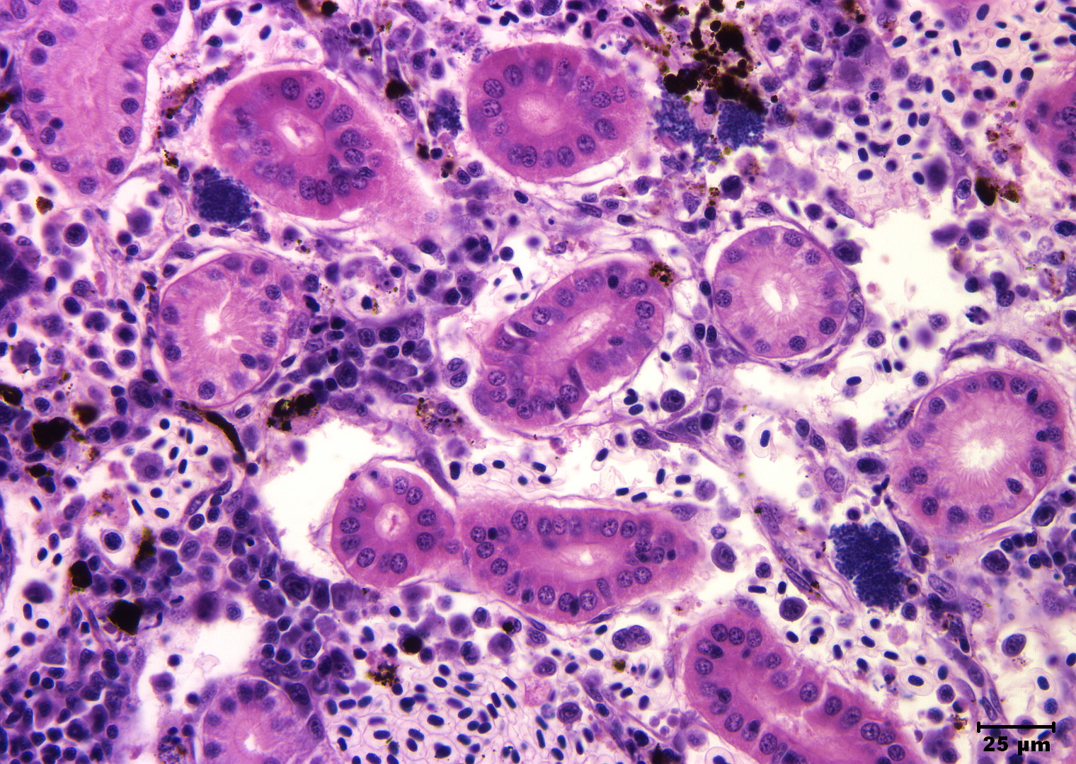
 | Slide # 18 Kidney Aeromonas salmonicida H&E 400X.jpg | All Pathogens | |
 | Slide # 58 Liver Mycobacteria H&E 400X.jpg | All Pathogens | |
 | database_SLH0028 - Double infection.jpg | All Pathogens, Fact Sheet Images | |
 | DigsFish_Zeylandicobdella-est-cod.jpg | All Pathogens, Contributed Images | estuary cod - Epinephelus coioides |
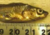
 | FishPathogens_LargescaleSucker_P1000426.jpg | All Pathogens | largescale sucker |
 | Slide # 86 Gills White Sturgeon Iridovirus H&E 400X 2.png | All Pathogens | white sturgeon |
 | BO_Loma_x63_19.jpg | All Pathogens | |
 | MYX_XX15_05b_x100nom_1shot.jpg | All Pathogens, Fact Sheet Images | |
 | 177_7781.JPG | All Pathogens, Fact Sheet Images | |
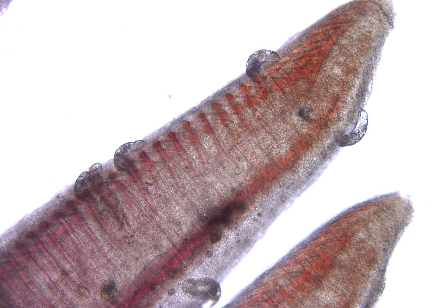
 | Dactylogyrus_01.jpg | All Pathogens, Fact Sheet Images | |
 | Cs2mo_poly3_02_x40bf_1h.jpg | All Pathogens, Fact Sheet Images | Manayunkia sp. polychaete worm |
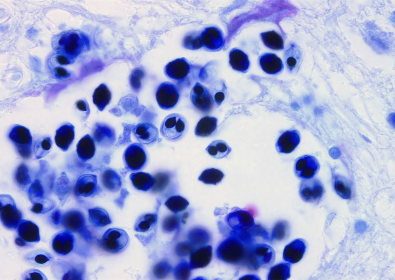
 | M.kisutchi_ed.jpg | All Pathogens, Fact Sheet Images | |
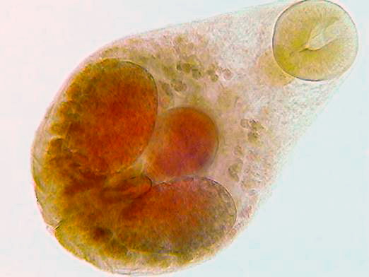
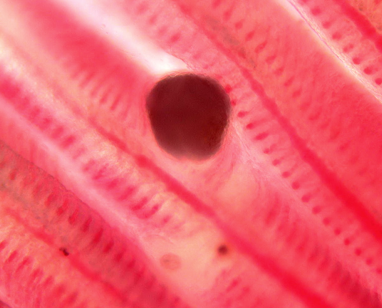
 | nanophyetus_salmonicola_CB01.jpg | All Pathogens, Fact Sheet Images | |
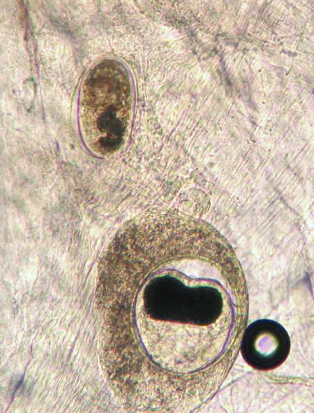
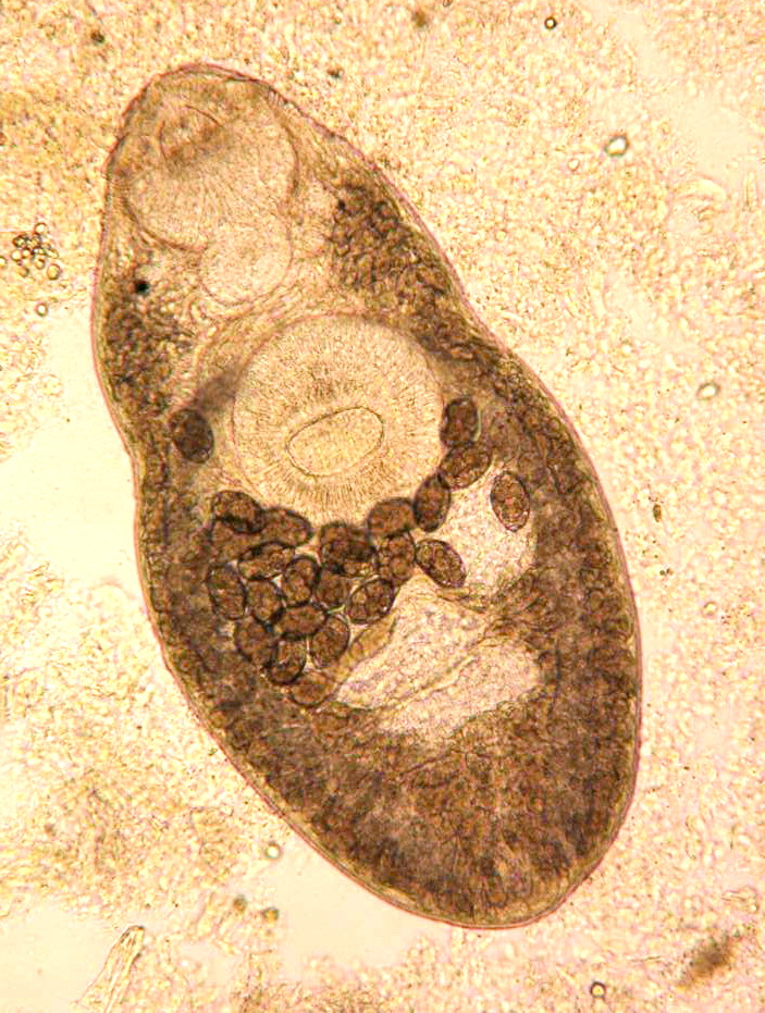
 | nanophyetus_salmonicola_CB08.jpg | All Pathogens, Fact Sheet Images | |
 | P1020307_annotated.jpg | All Pathogens, Fact Sheet Images | bluegill |
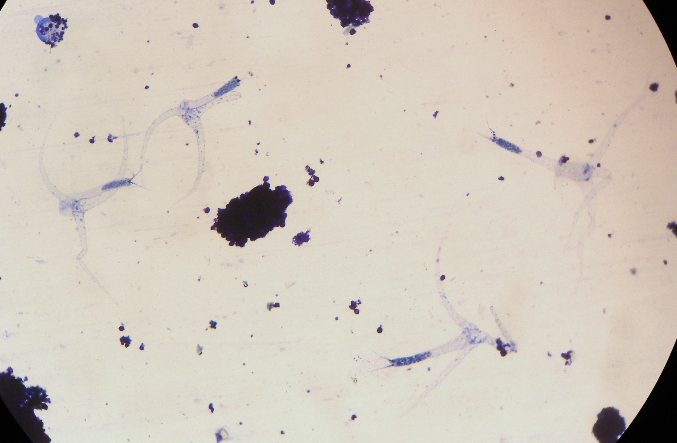
 | glochidia_CB05.jpg | All Pathogens | |
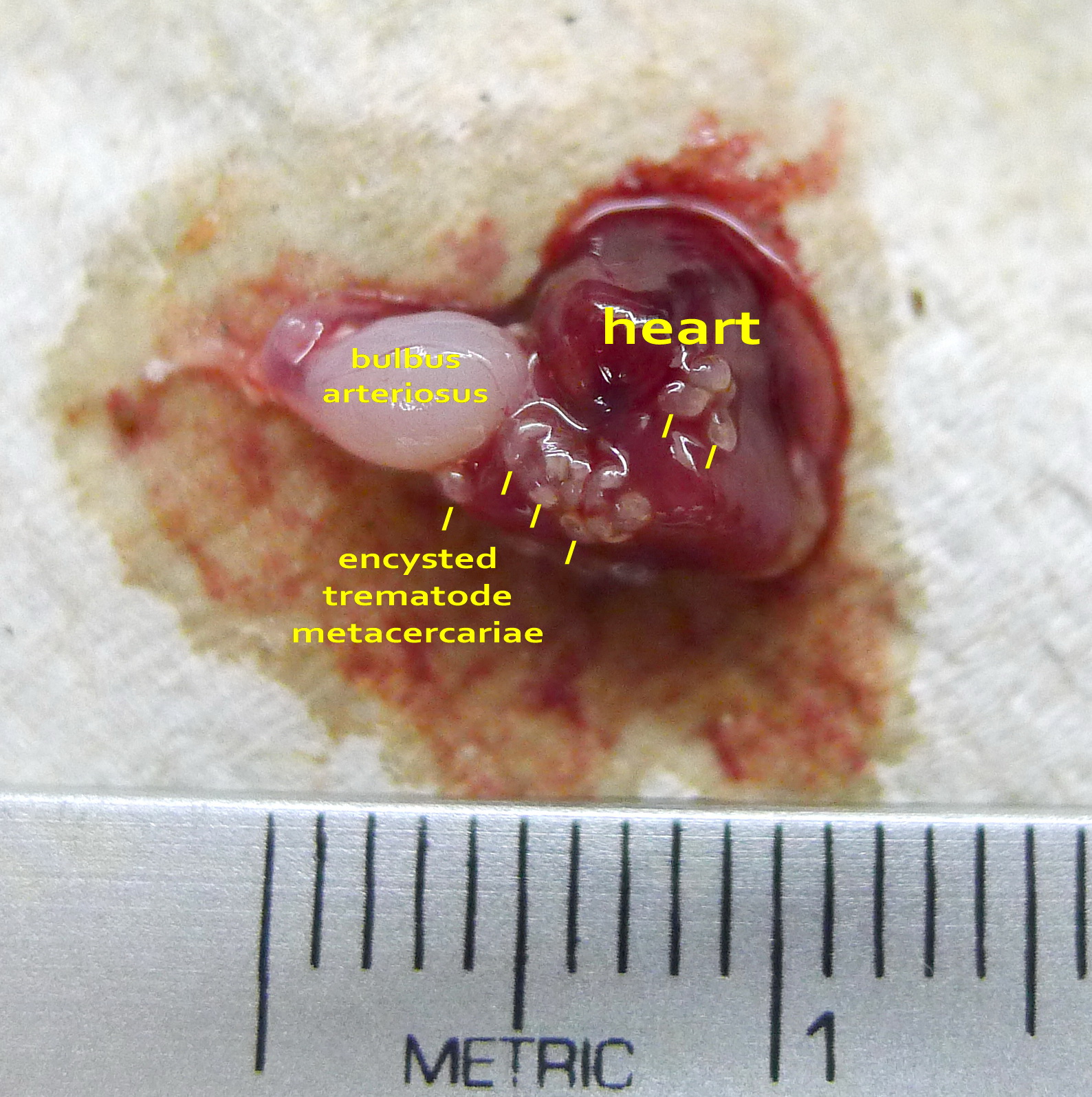
 | trematode_unidentified_inGB_CB04.jpg | All Pathogens, Fact Sheet Images | |
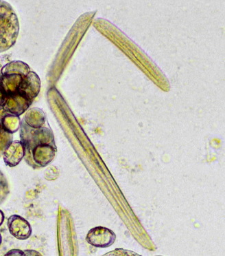
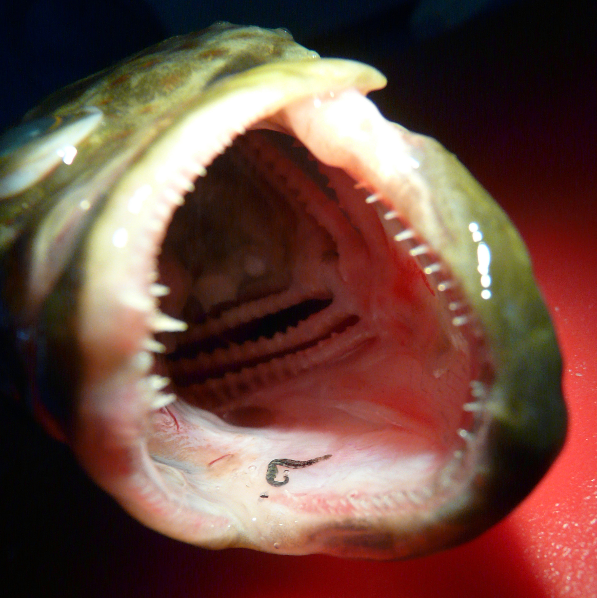
 | nematode_Camallanus_CB01.jpg | All Pathogens, Fact Sheet Images | |
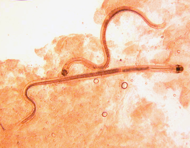
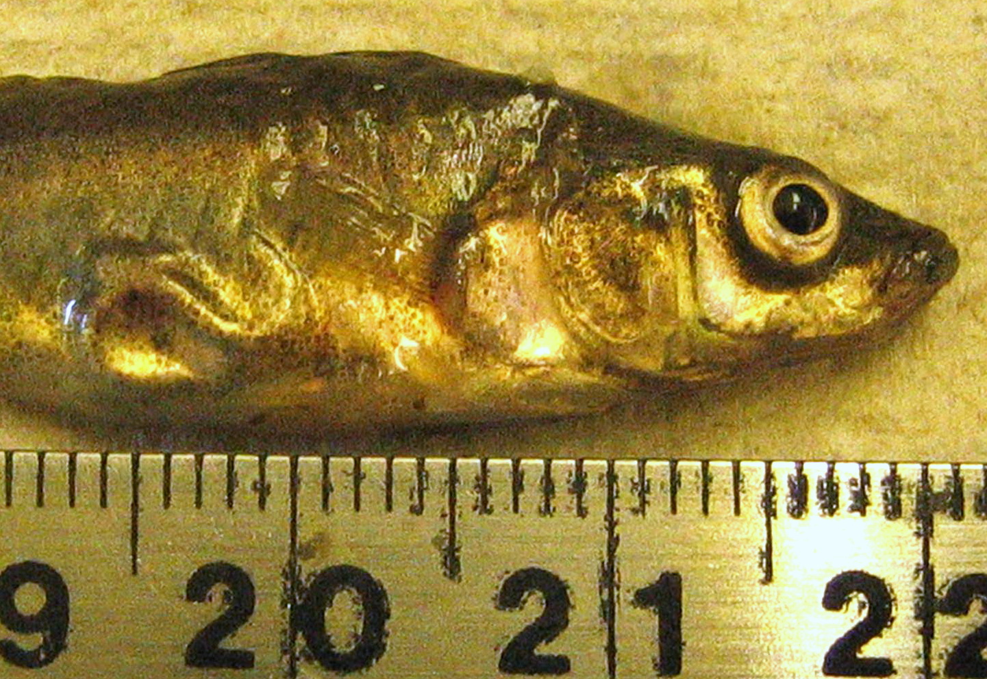
 | nematode_Eustrongylides_CB01.jpg | All Pathogens, Fact Sheet Images | threespine stickleback |