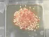
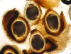
 | Glochidia_02.jpg | All Pathogens, Fact Sheet Images | |
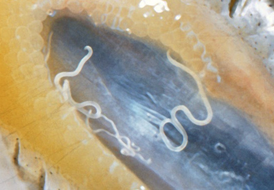
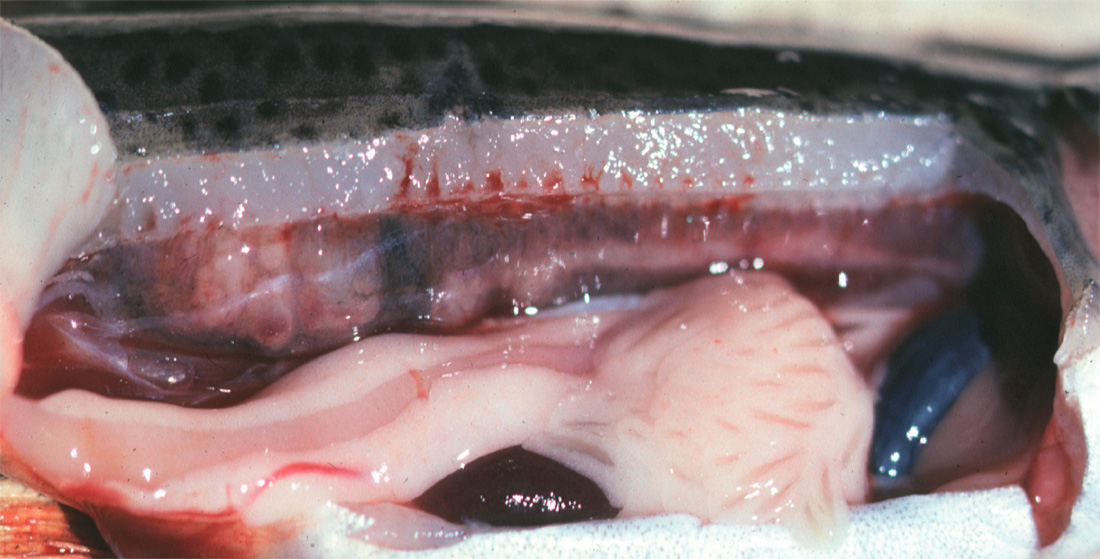
 | nematode_Philonema_spp_CB02.jpg | All Pathogens, Fact Sheet Images | |
 | nematode_Capillaria_CB02.jpg | All Pathogens, Fact Sheet Images | |
 | nematode_Anasakis_sp_CB01.jpg | All Pathogens, Fact Sheet Images | |
 | Acanthocephala_CB02.jpg | All Pathogens, Fact Sheet Images | |
 | Acanthocephala_Echinorhynchus_lageniformis_CB03.jpg | All Pathogens, Fact Sheet Images | |
 | Acanthocephala_Neoechinorhynchus_sp_CB05.jpg | All Pathogens, Fact Sheet Images | |
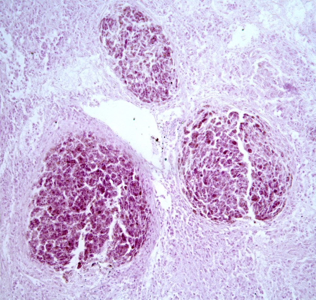
 | trematode_Posthodiplostomum_minimum_CB03.jpg | All Pathogens, Fact Sheet Images | |
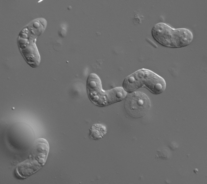
 | hemoflagellate_Cryptobia_salmositica_CB03.jpg | All Pathogens, Fact Sheet Images | |
 | nematode_Anasakis_simplex_CB01.jpg | All Pathogens, Fact Sheet Images | |
 | nitzschia_CB01.jpg | All Pathogens, Fact Sheet Images | white sturgeon |
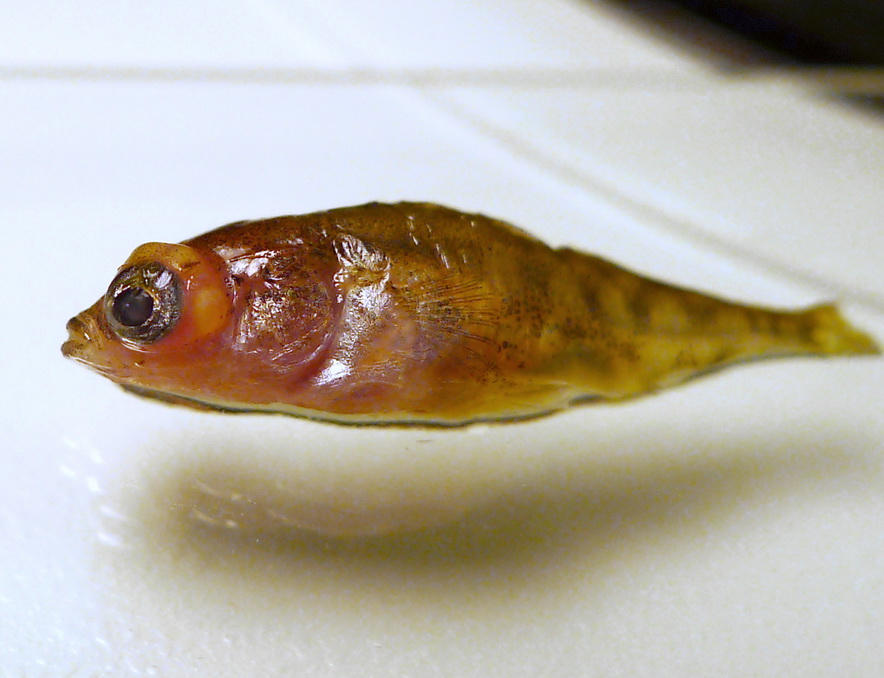
 | P1060630_diplostomum.jpg | All Pathogens | threespine stickleback |
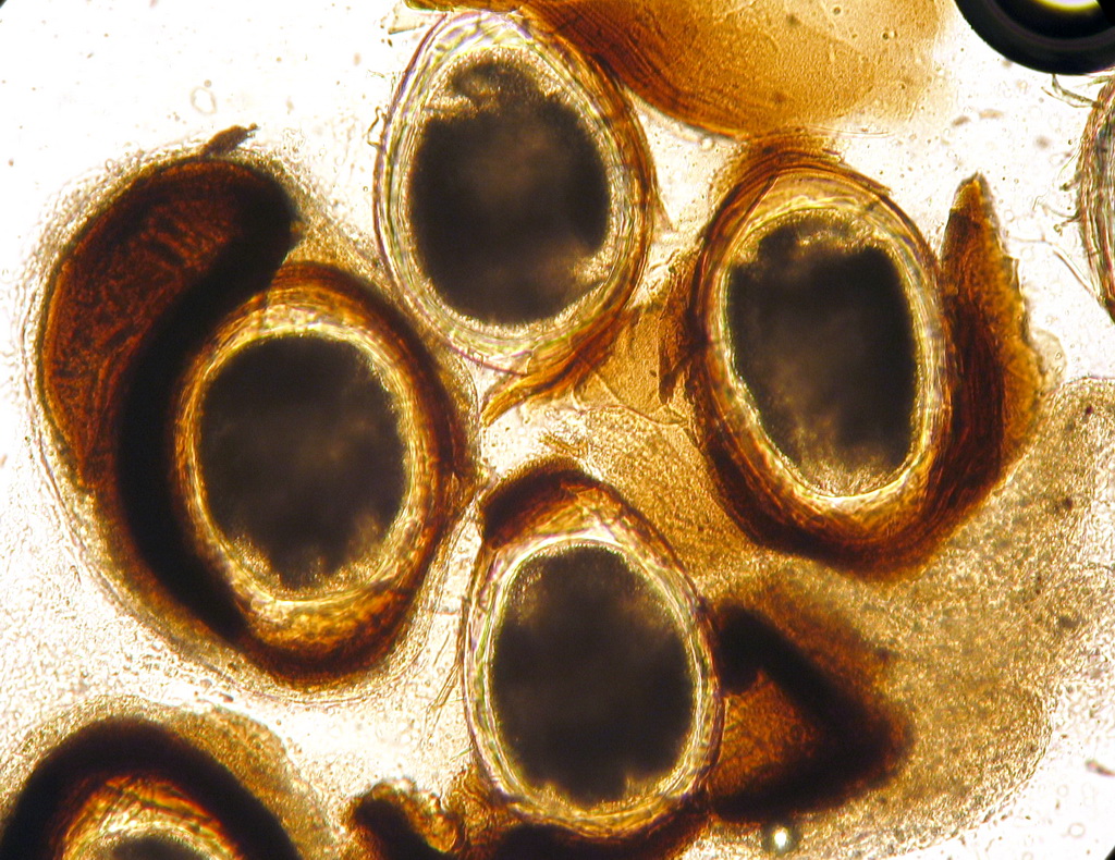
 | CB_146_4665.jpg | All Pathogens, Fact Sheet Images | threespine stickleback |
 | CB_GFFR043.jpg | All Pathogens, Fact Sheet Images | goldfish |
 | WhirlingDisease_DSC03051.jpg | All Pathogens | rainbow trout |
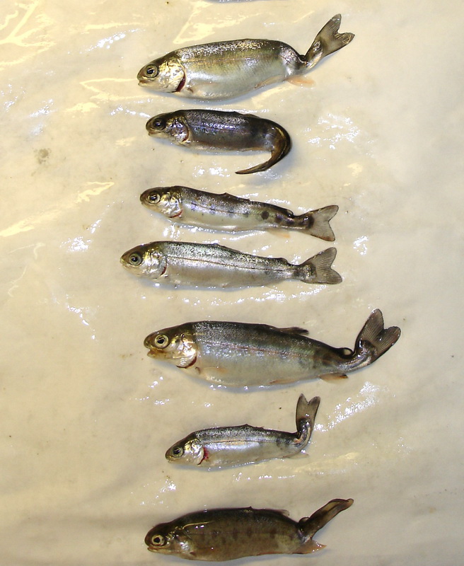
 | IMG_20130402_rbtCs2.jpg | All Pathogens | rainbow trout |
 | FishPath_placeholder.gif | All Pathogens | |
 | FishPathogens_Cryptobia_IMG_8231.jpg | All Pathogens, Fact Sheet Images | |
 | JJ0004-SAed_cystidicola_cropped.jpg | All Pathogens | |
 | Renibacterium_3.jpg | All Pathogens | |
 | BO_myco_skulpin_x20_03.jpg | All Pathogens | sculpin (unspecified) |
 | L14m7809_0900a_100nm_1_12a.jpg | All Pathogens, Fact Sheet Images | |
 | Myxobolus_squamalis_under_scale_2.jpg | All Pathogens | |
 | white_grub_02.jpg | All Pathogens, Fact Sheet Images | |
 | CB_IMG_5197.jpg | All Pathogens, Fact Sheet Images | |
 | Ichthyophonus1ed.jpg | All Pathogens, Fact Sheet Images | |
 | diplostomum_CB04.jpg | All Pathogens | threespine stickleback |
 | nanophyetus_salmonicola_CB06.jpg | All Pathogens, Fact Sheet Images | |
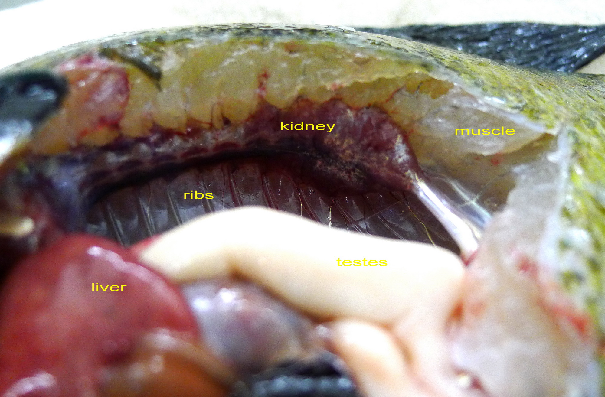
 | P1020305_annotated.jpg | All Pathogens, Fact Sheet Images | bluegill |
 | cesode_Proteocephalus_sp_CB01.jpg | All Pathogens, Fact Sheet Images | |