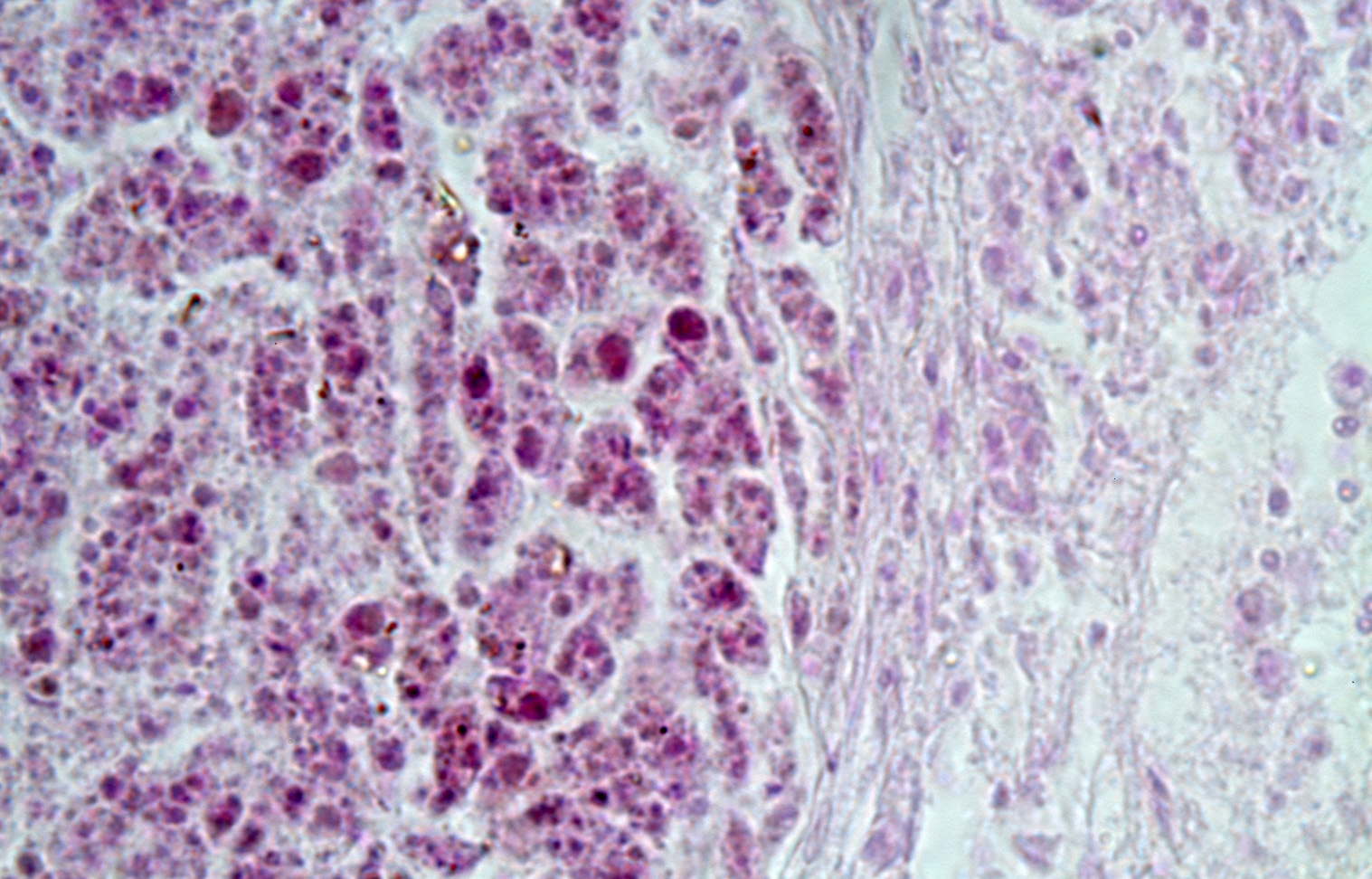
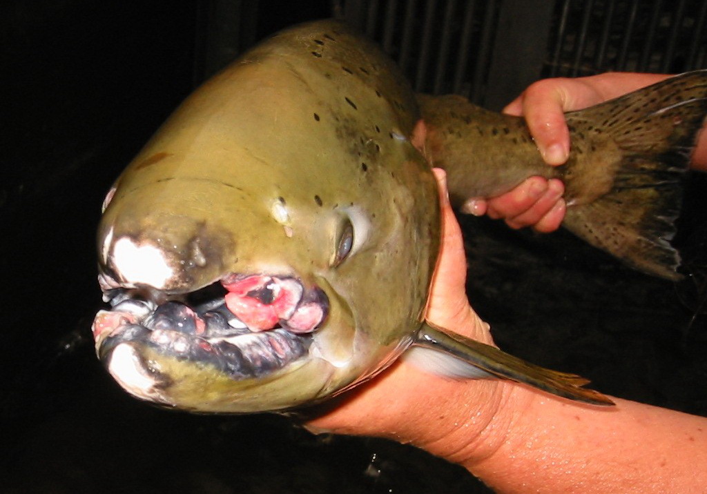
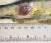
 | JJ0009_SAed.jpg | All Pathogens | |
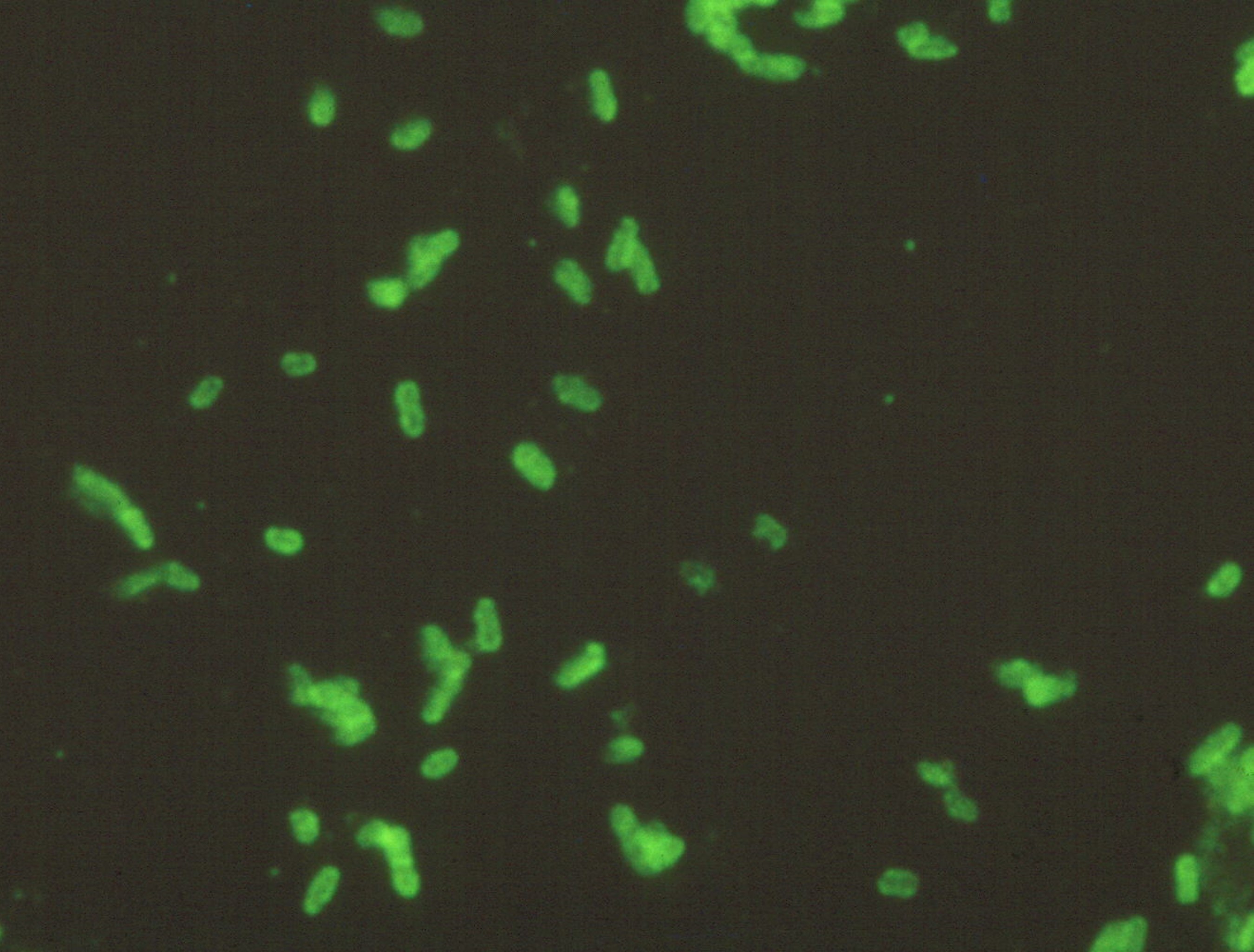
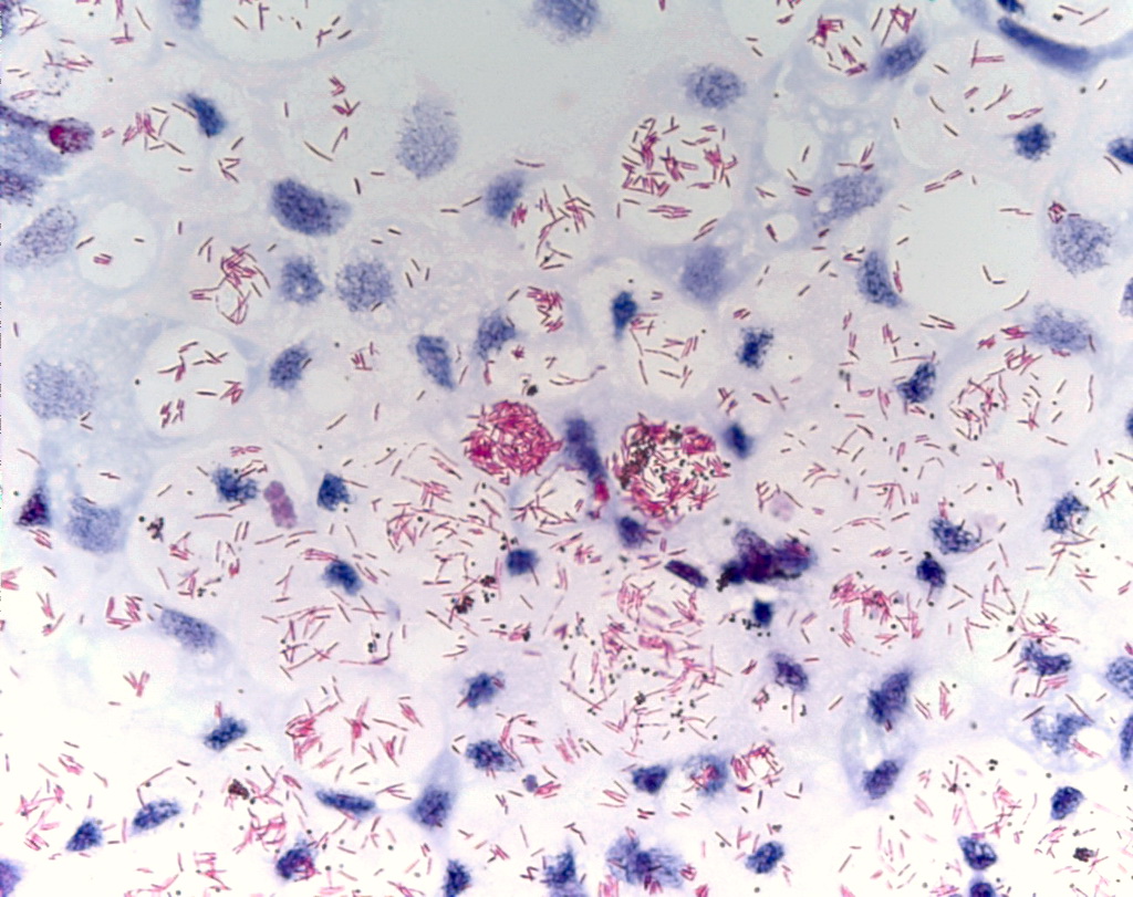
 | Renibacterium_1.jpg | All Pathogens | |
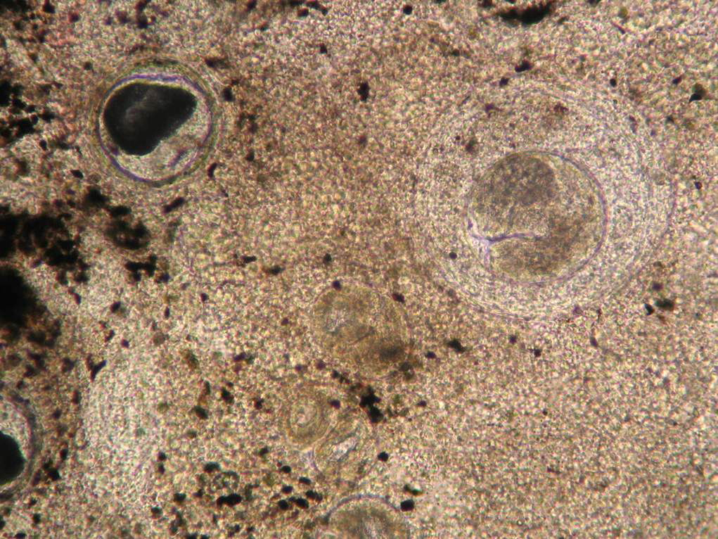
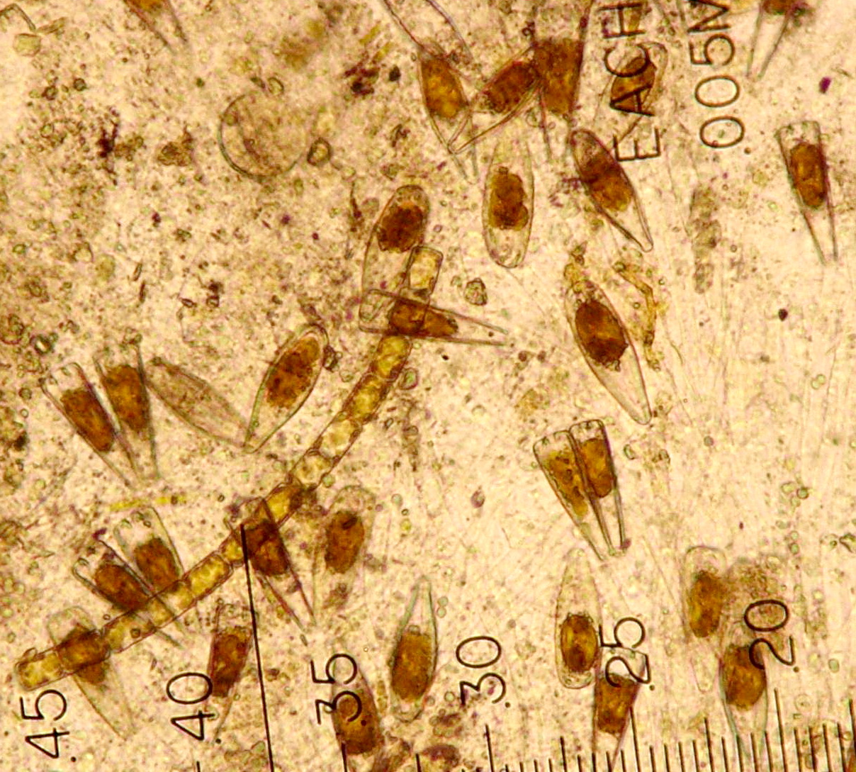
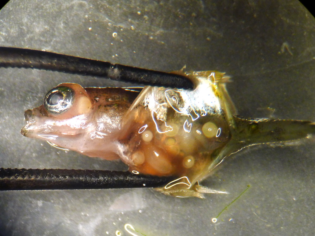
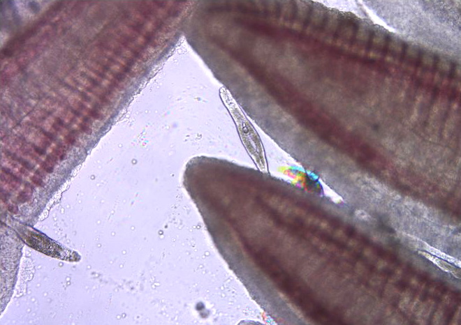
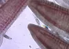
 | BO_myco_skulpin_x100_04.jpg | All Pathogens | sculpin (unspecified) |
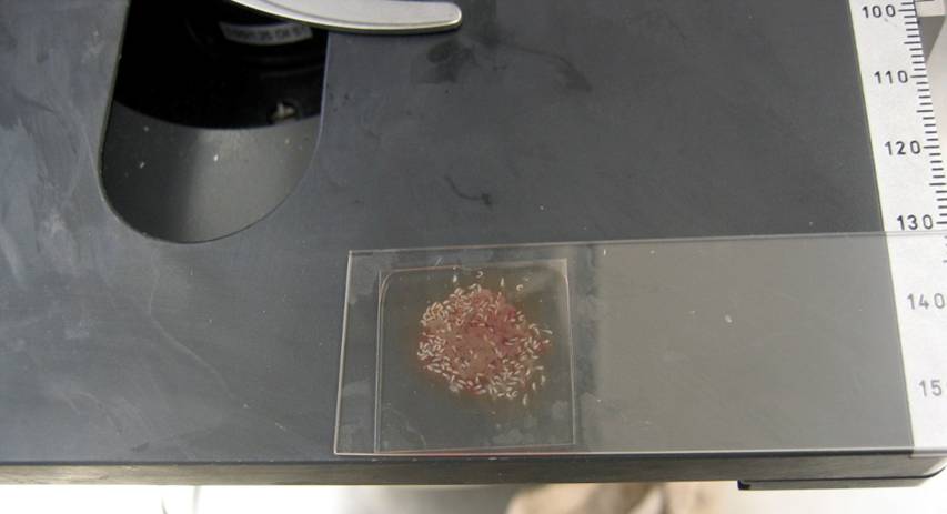
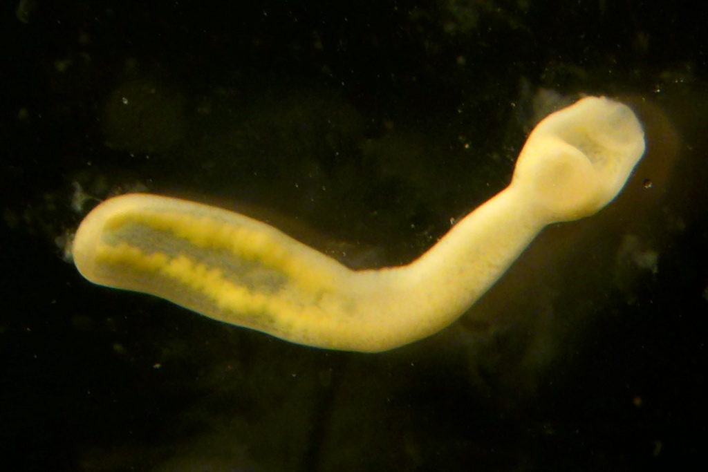
 | Clinostomum_05.jpg | All Pathogens, Fact Sheet Images | |
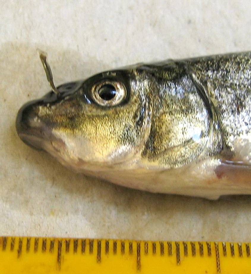
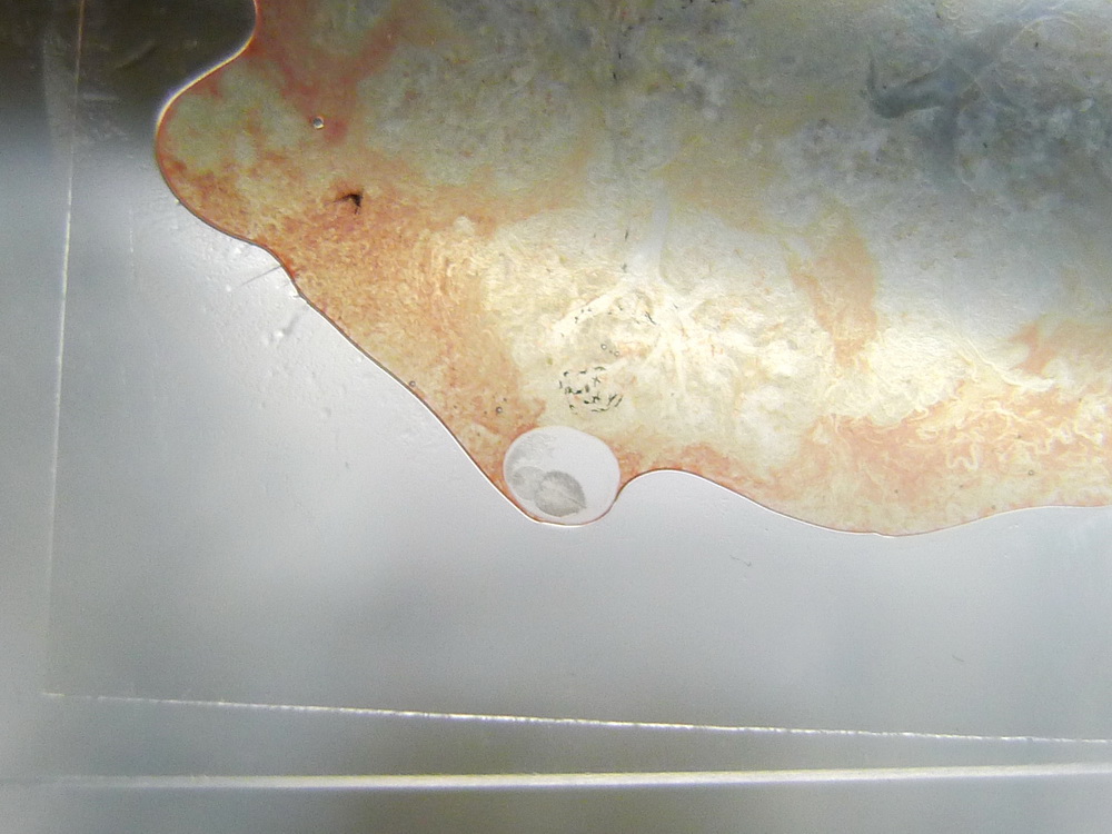
 | Myxobolus_squamalis_puckered_scales.jpg | All Pathogens, Fact Sheet Images | |
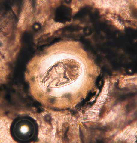
 | trematode_metacercaria_family_Heterophyidae_02.jpg | All Pathogens, Fact Sheet Images | |
 | apophallus_CB01.jpg | All Pathogens, Fact Sheet Images | |
 | epistylis_CB01.jpg | All Pathogens, Fact Sheet Images | |
 | lernea_CB01.jpg | All Pathogens, Fact Sheet Images | |
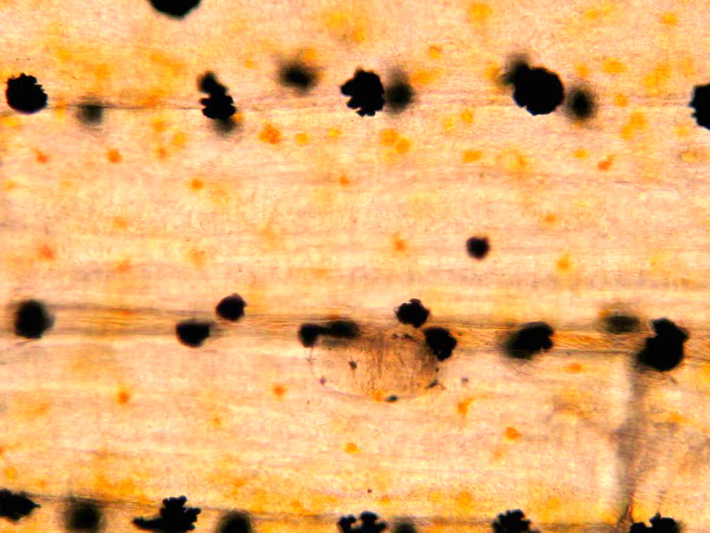
 | neascus_CB01.jpg | All Pathogens, Fact Sheet Images | |
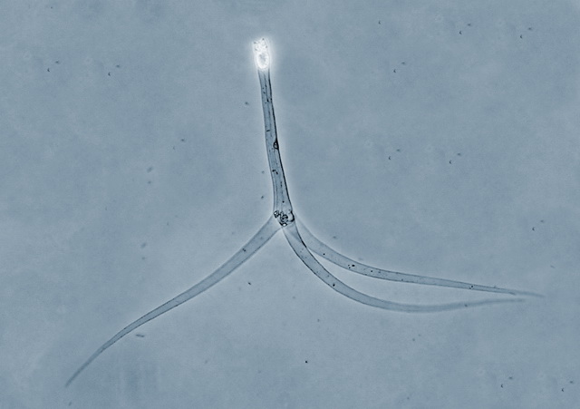
 | sanguinicola_CB07.jpg | All Pathogens, Fact Sheet Images | |
 | P1020572crop.jpg | All Pathogens | goldfish |
 | cesode_Proteocephalus_sp_CB01.jpg | All Pathogens, Fact Sheet Images | |
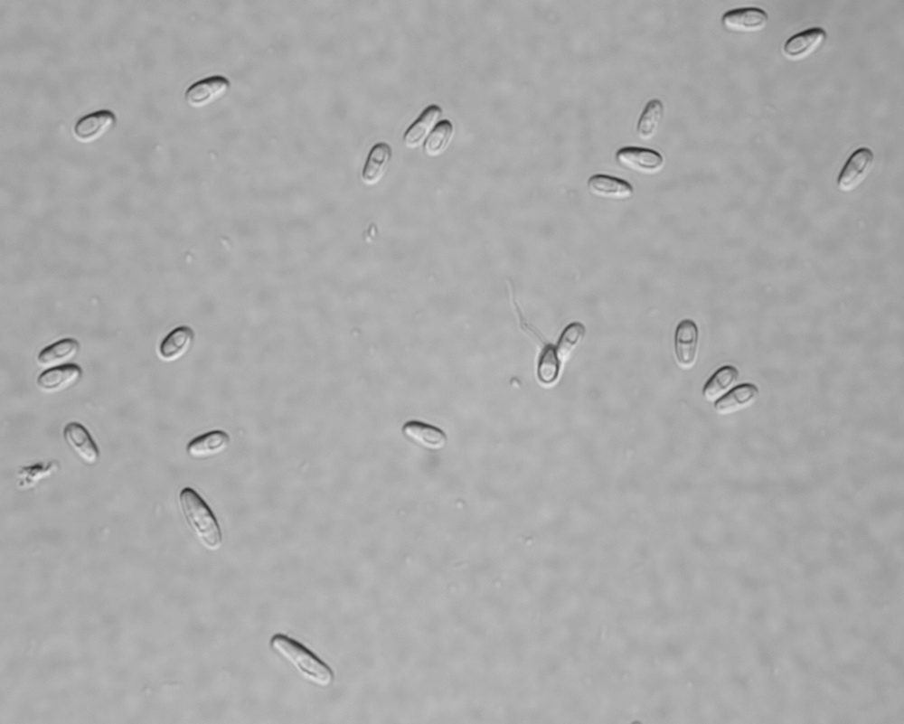
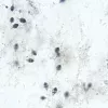
 | SA_Generic_TAM.jpg | All Pathogens, Fact Sheet Images | |
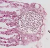
 | SA_MYXCC02_02_x100.jpg | All Pathogens, Fact Sheet Images | sculpin (unspecified) |
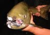
 | P1020180_SA_whitegrub.jpg | All Pathogens, Fact Sheet Images | smallmouth bass |
 | P1060631_diplostomum.jpg | All Pathogens, Fact Sheet Images | threespine stickleback |
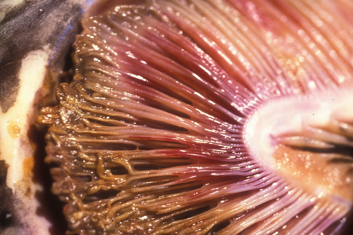
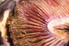 | RH_Fcolmngill.jpg | All Pathogens, Fact Sheet Images | Chinook salmon |
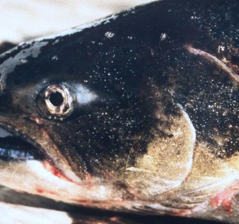
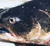 | CB_adult_Ich_ChF.jpg | All Pathogens, Fact Sheet Images, Top 10 | Fall Chinook salmon |
 | CB_Kla_ModCWD12.jpg | All Pathogens, Fact Sheet Images | |
 | CB_MYCORASB.jpg | All Pathogens, Fact Sheet Images | |
 | CB_ChS_tumors3.jpg | All Pathogens, Fact Sheet Images | Spring Chinook salmon |
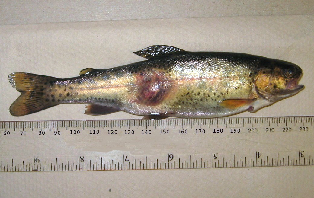
 | WhirlingDisease_DSC03056.jpg | All Pathogens | rainbow trout |
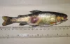
 | P1000380ed.jpg | All Pathogens, Fact Sheet Images | smallmouth bass |
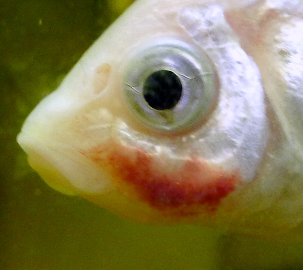
 | CB_Aeromonas_01.jpg | All Pathogens, Fact Sheet Images | rainbow trout |
 | CB_Dactylogyrus01.jpg | All Pathogens, Fact Sheet Images | |
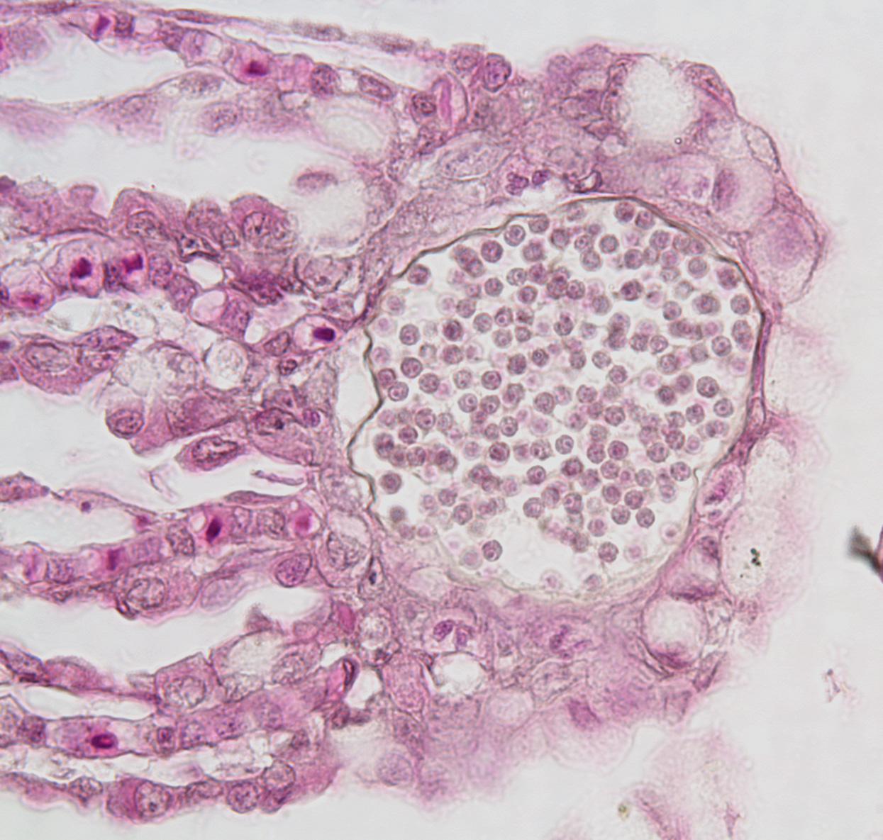
 | BO_Dermocystidium_x63_23.jpg | All Pathogens, Fact Sheet Images | |
 | BO_Hexamita_x63_34.jpg | All Pathogens, Fact Sheet Images | |
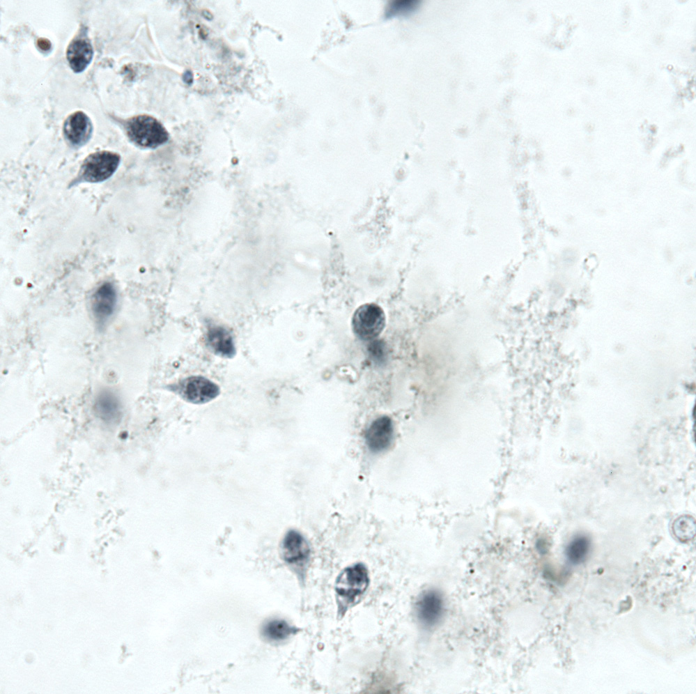
 | BO_Ichthyobodo_x63_32.jpg | All Pathogens, Fact Sheet Images | Chinook salmon |
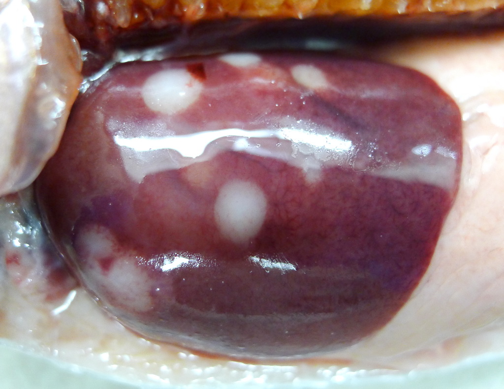
 | P1080678_Cshasta_liver.jpg | All Pathogens | rainbow trout |