 | Renibacterium_1.jpg | All Pathogens | |
 | BO_myco_skulpin_x100_04.jpg | All Pathogens | sculpin (unspecified) |
 | 20120911e_07a_x100nm1h.jpg | All Pathogens, Fact Sheet Images | redside shiner |
 | L14m7709_1800b_100bf_1_3a.jpg | All Pathogens, Fact Sheet Images | |
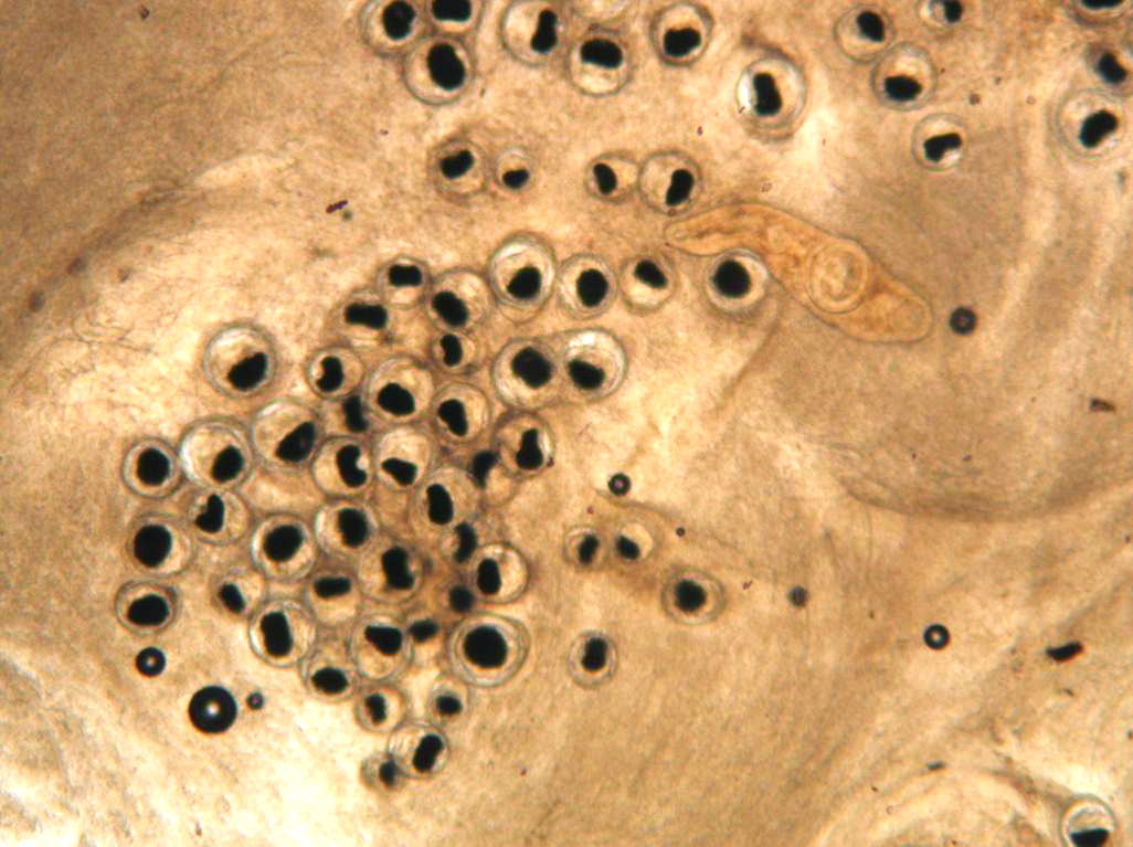
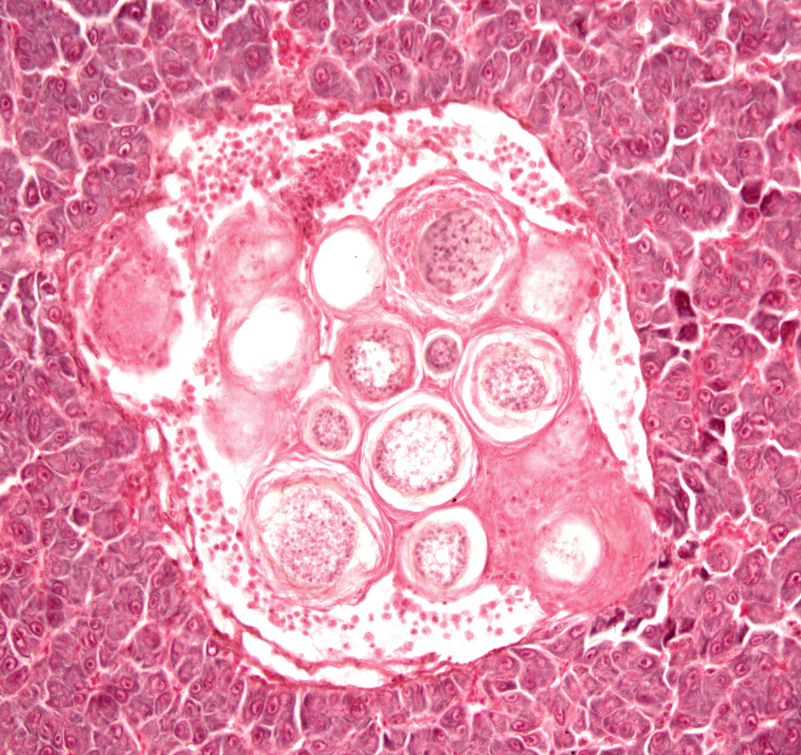
 | Henneguya_salminicola_cysts_02_crop.jpg | All Pathogens, Fact Sheet Images | |
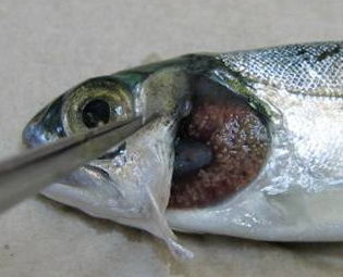
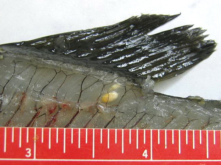
 | Salmincola_03.jpg | All Pathogens, Fact Sheet Images | |
 | ichtyoph.jpg | All Pathogens, Fact Sheet Images | |
 | nanophyetus_salmonicola_CB07.jpg | All Pathogens, Fact Sheet Images | |
 | P1020306.jpg | All Pathogens, Fact Sheet Images | bluegill |
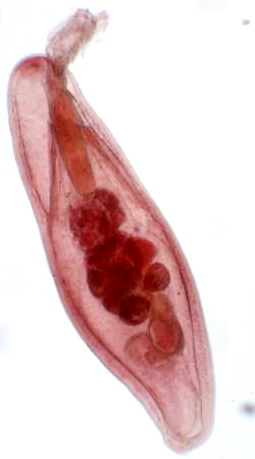
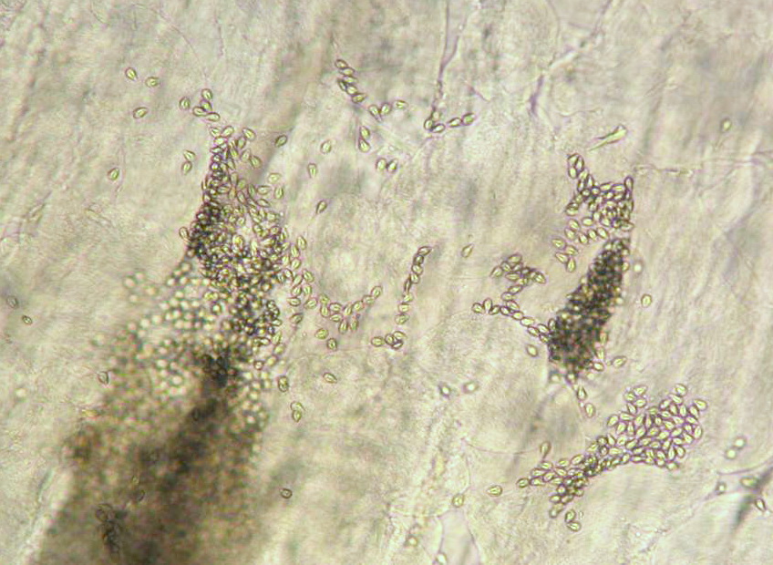
 | Glochidia_01.jpg | All Pathogens, Fact Sheet Images | |
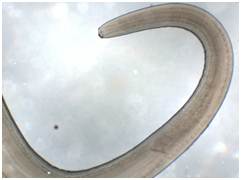
 | nematode_Philonema_spp_CB01.jpg | All Pathogens, Fact Sheet Images | |
 | nematode_Capillaria_CB03.jpg | All Pathogens, Fact Sheet Images | |
 | nematode_Salvelinema_walkeri_CB04.jpg | All Pathogens, Fact Sheet Images | |
 | Acanthocephala_CB03.jpg | All Pathogens, Fact Sheet Images | |
 | Acanthocephala_Echinorhynchus_lageniformis_CB01.jpg | All Pathogens, Fact Sheet Images | |
 | Acanthocephala_Neoechinorhynchus_sp_CB06.jpg | All Pathogens, Fact Sheet Images | |
 | trematode_Clinostomum_marginatum_CB03.jpg | All Pathogens, Fact Sheet Images | |
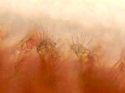
 | tricophyra_CB01.jpg | All Pathogens, Fact Sheet Images | |
 | nematode_Anasakis_simplex_CB02.jpg | All Pathogens, Fact Sheet Images | |
 | Minsidiosus_CB02.jpg | All Pathogens, Fact Sheet Images | |
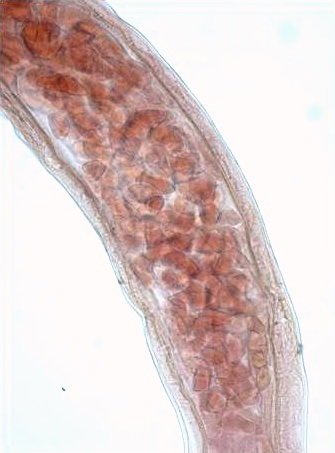
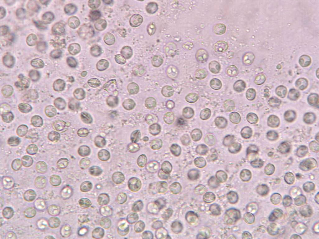
 | SA_Loma_01a_x10bf_9shot.jpg | All Pathogens, Fact Sheet Images | |
 | CB_BlacSptSucker.jpg | All Pathogens, Fact Sheet Images | sucker (unidentified) |
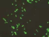
 | CB_Amoeba19.jpg | All Pathogens, Fact Sheet Images | |
 | SLH_DSC00179.jpg | All Pathogens, Top 10 | rainbow trout |
 | FishPath_placeholder.gif | All Pathogens | |
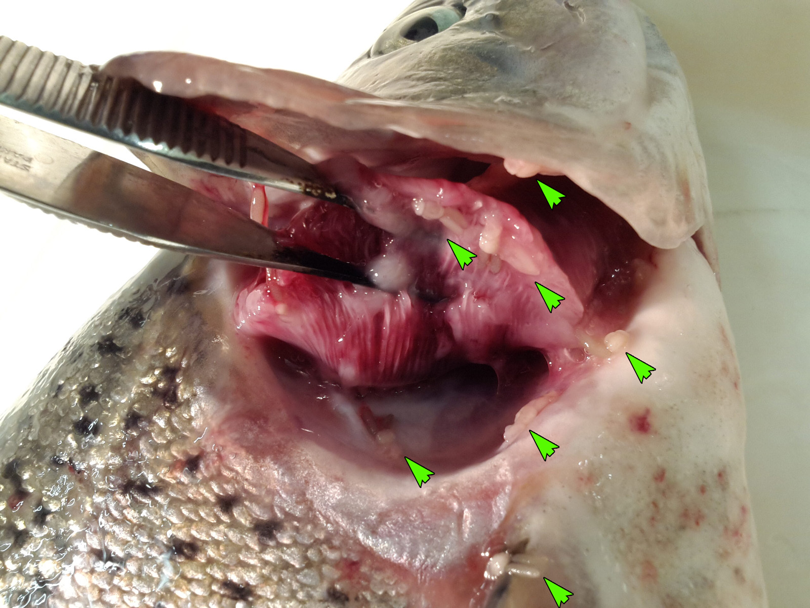
 | FishPathogens_Salmincola_IMG_20140421_201935.jpg | All Pathogens | rainbow trout |
 | JJ0043_SAed_EIBS.jpg | All Pathogens | |
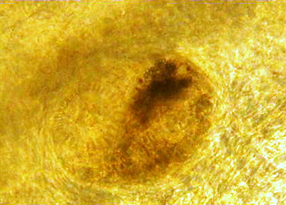
 | BO_Ichthyophonus_x20_37.jpg | All Pathogens | |
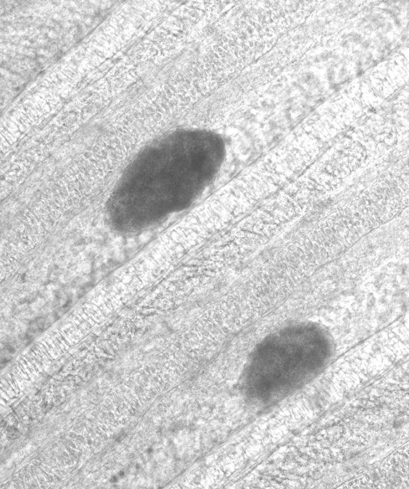
 | Myxobolus_squamalis_cyst.jpg | All Pathogens, Fact Sheet Images | |
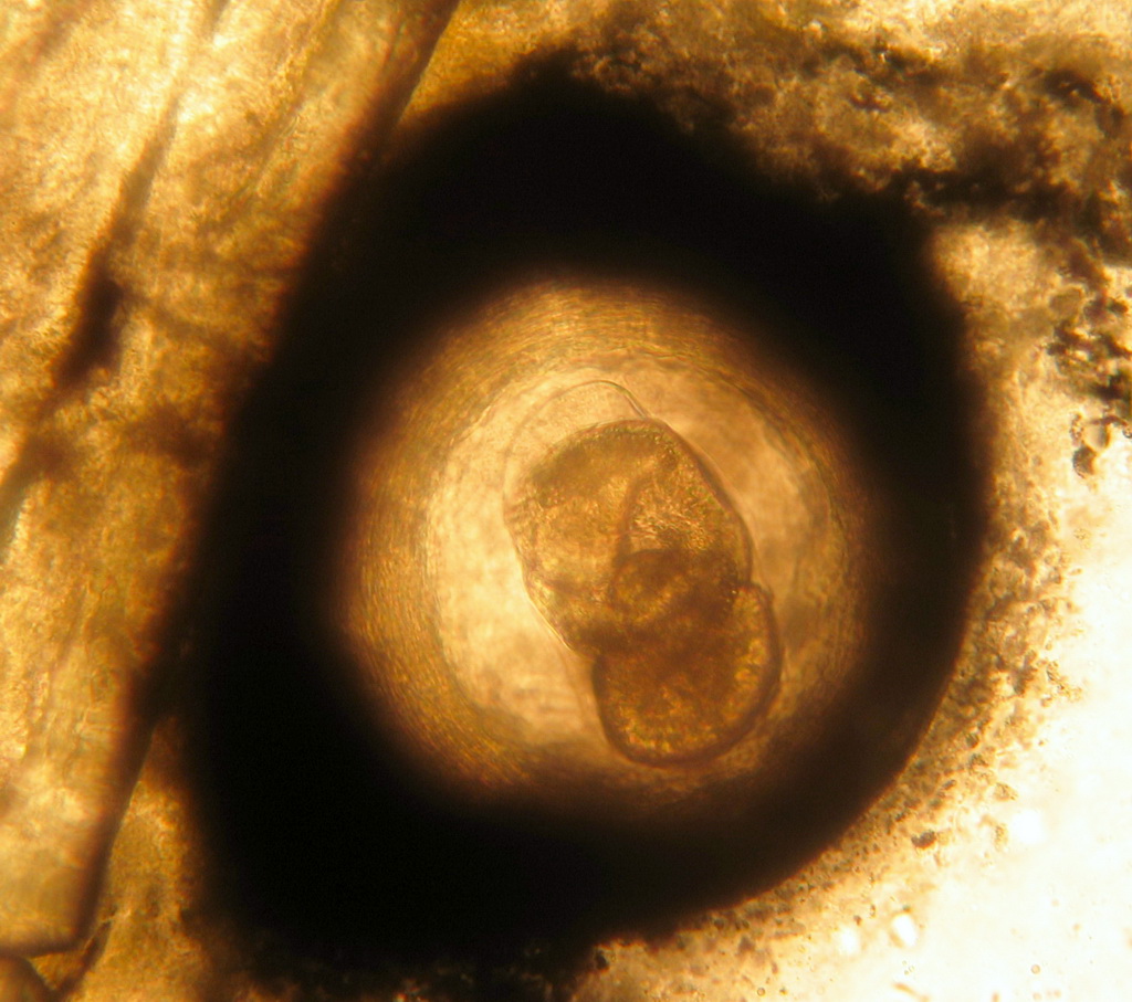
 | trematode_metacercaria_family_Heterophyidae_03.jpg | All Pathogens, Fact Sheet Images | |