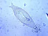
| chilinodella_CB01.jpg | All Pathogens, Fact Sheet Images | |
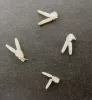
 | gyrodactylus_CB01.jpg | All Pathogens, Fact Sheet Images | |
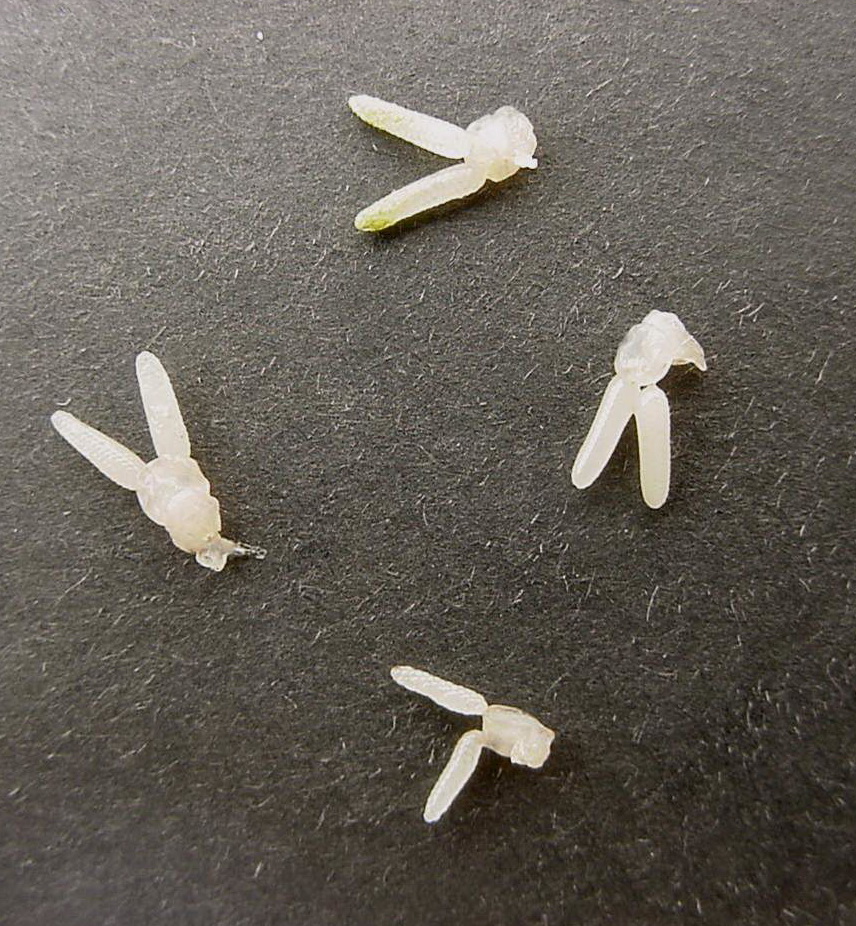
 | lernea_CB02.jpg | All Pathogens | |
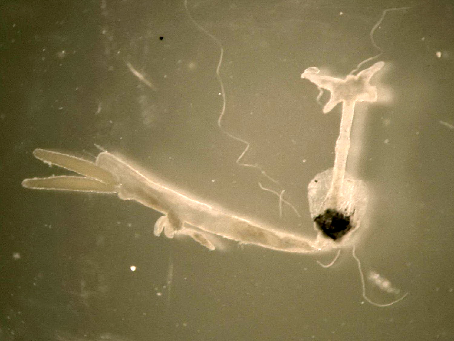
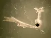
 | salmincola-CB04.jpg | All Pathogens, Fact Sheet Images | |
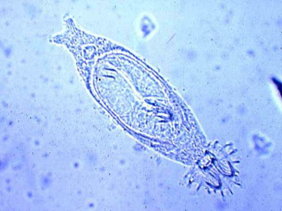
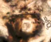 | sanguinicola_CB08.jpg | All Pathogens, Fact Sheet Images | |
 | cesode_Proteocephalus_sp_CB02.jpg | All Pathogens, Fact Sheet Images | |
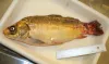 | SA_IMG_4724.jpg | All Pathogens, Hosts and other organisms | common carp |
 | SA_157_5710.jpg | All Pathogens, Fact Sheet Images | sculpin (unspecified) |
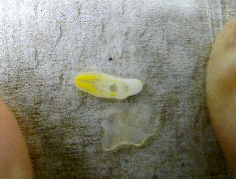
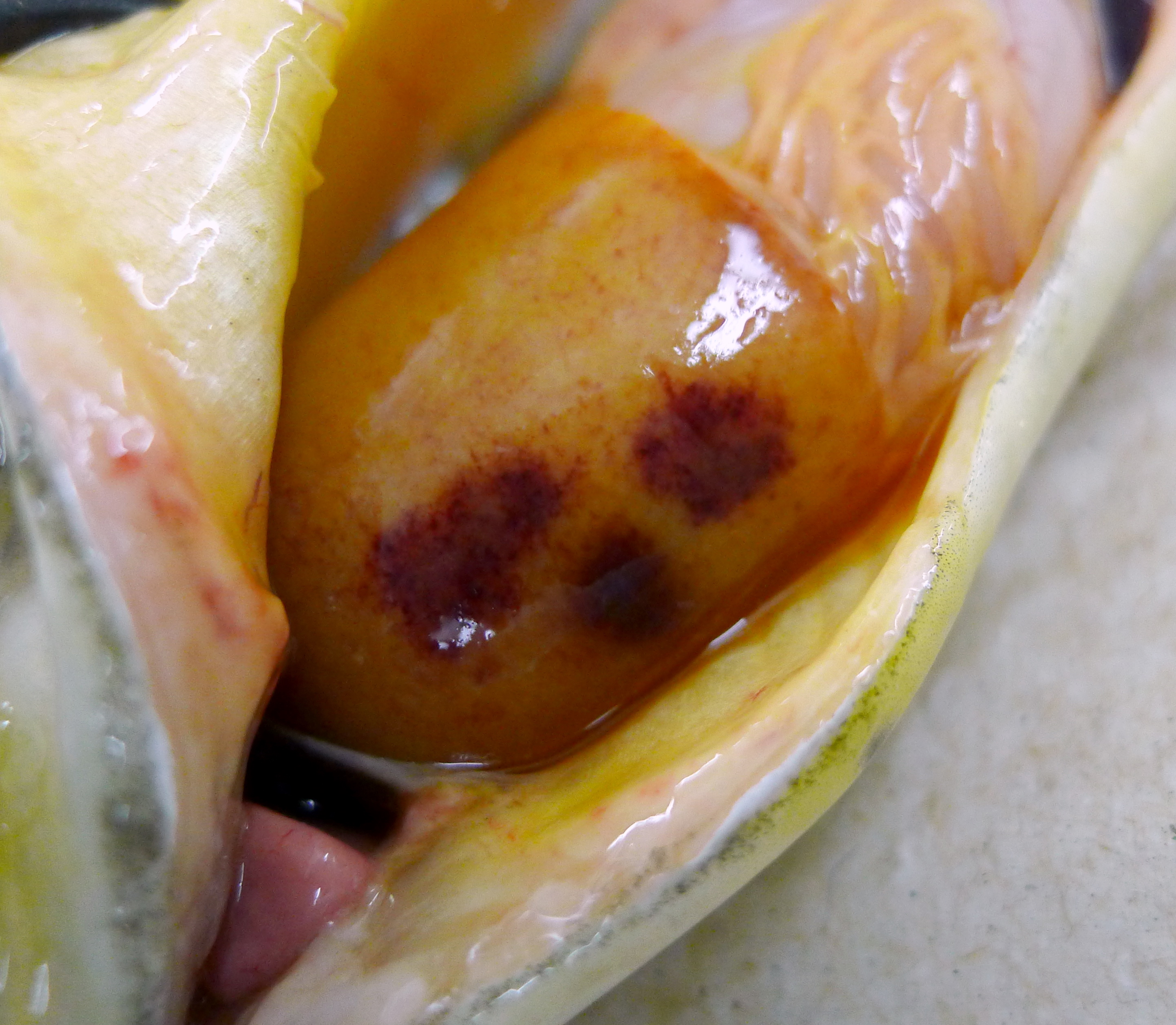
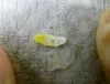 | SA_P1020178_yellowgrub.jpg | All Pathogens, Fact Sheet Images | smallmouth bass |
 | P1020180_SA_zoomed.jpg | All Pathogens, Fact Sheet Images | smallmouth bass |
 | P1060624_diplostomum.jpg | All Pathogens | threespine stickleback |
 | CB_IMG_5012.jpg | All Pathogens, Fact Sheet Images | |
 | CB_HeavyIch.jpg | All Pathogens, Fact Sheet Images | |
 | CB_Myxobolus_sp5.jpg | All Pathogens, Fact Sheet Images | |
 | CB_ChS_Tumor_lips.jpg | All Pathogens, Fact Sheet Images | Spring Chinook salmon |
 | P1000377ed.jpg | All Pathogens, Fact Sheet Images | smallmouth bass |
 | CB_BKD_01.jpg | All Pathogens, Fact Sheet Images | |
 | VHSV_kaufman.jpg | All Pathogens, Fact Sheet Images | |
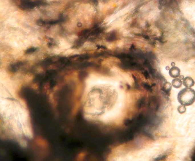
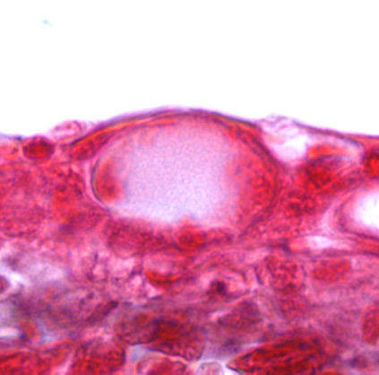
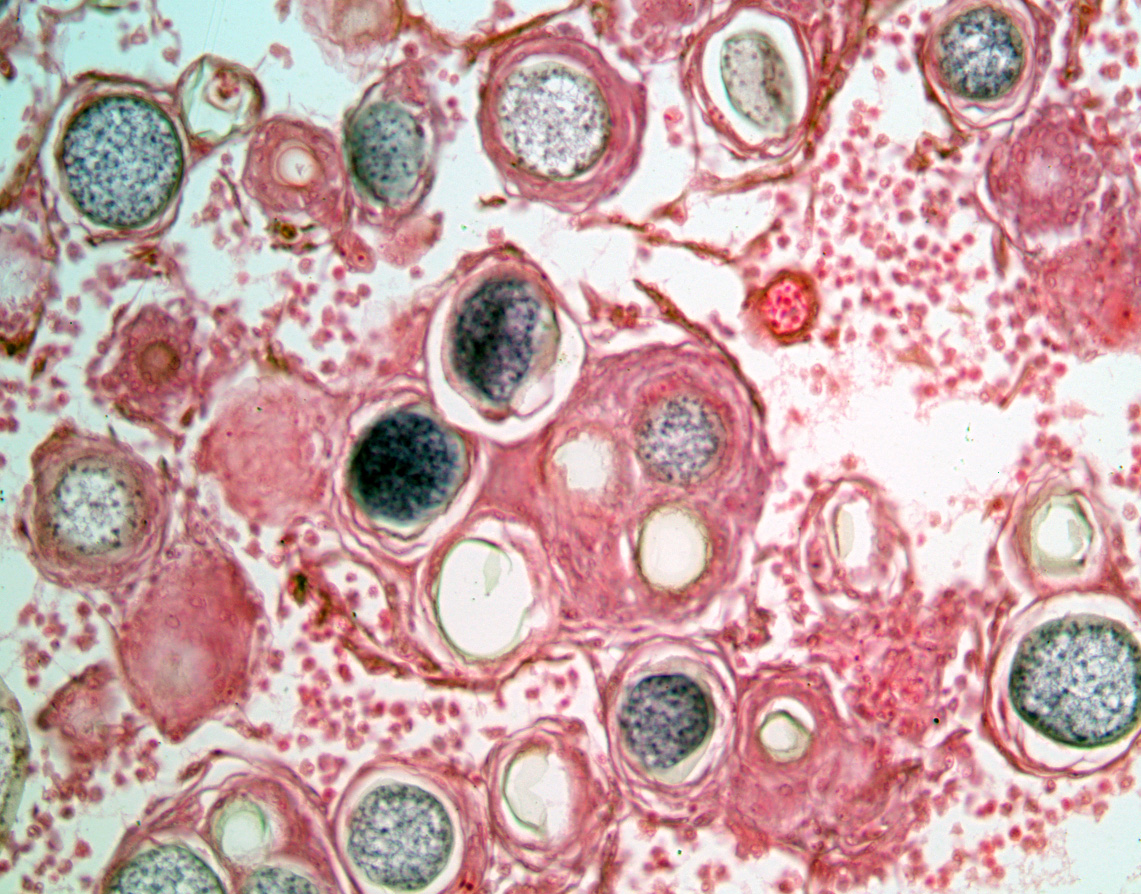
 | BO_Dermocystidium_x63_24.jpg | All Pathogens, Fact Sheet Images | |
 | BO_Hexamita_x63_35.jpg | All Pathogens, Fact Sheet Images | |
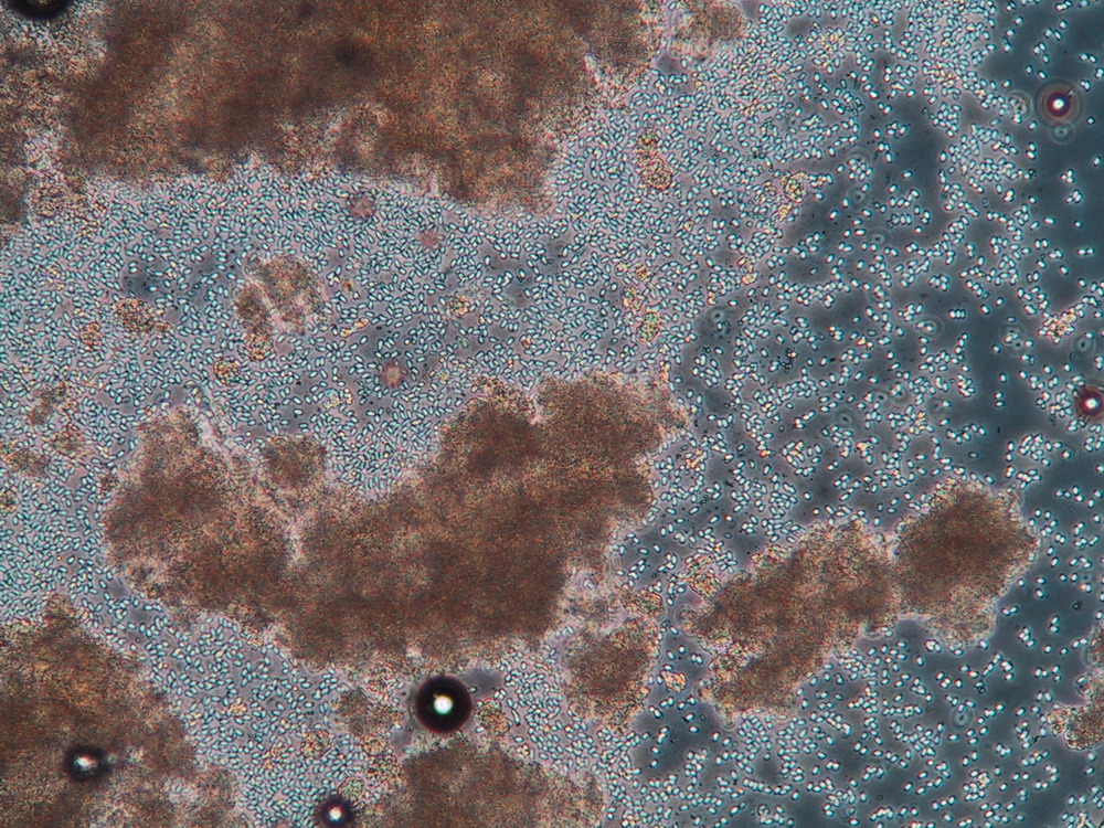
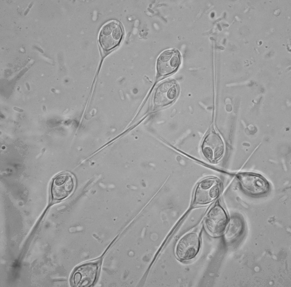
 | BO_Ichthyobodo_x63_33.jpg | All Pathogens, Fact Sheet Images | Chinook salmon |
 | P1070955_Cshasta.jpg | All Pathogens | rainbow trout |
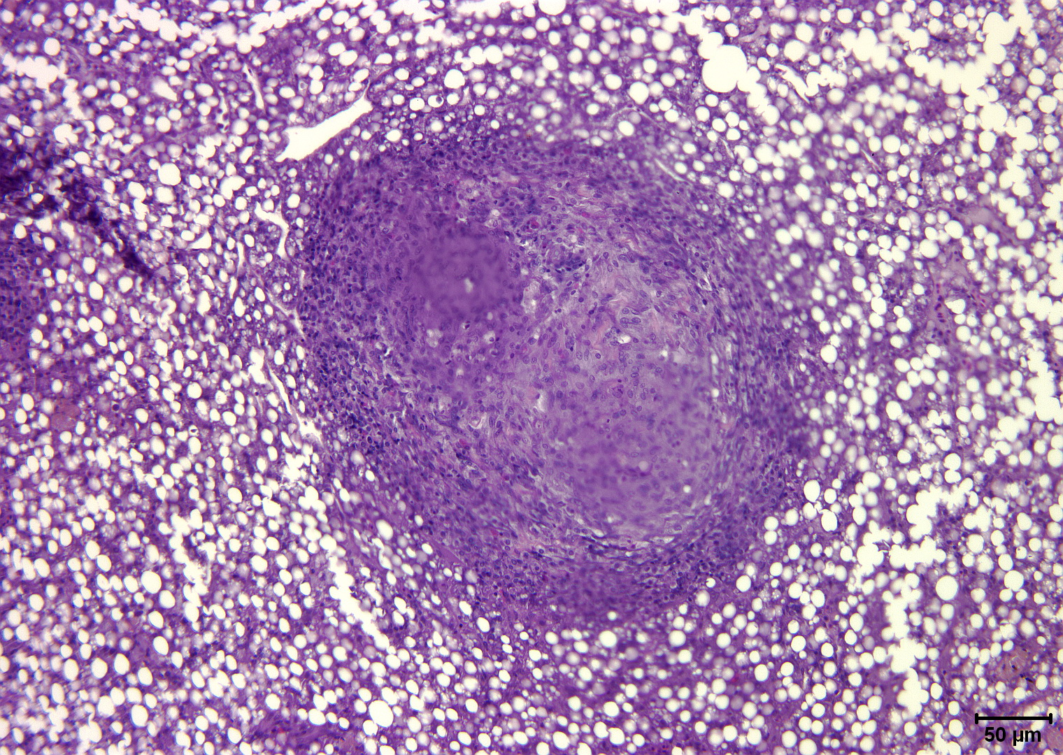
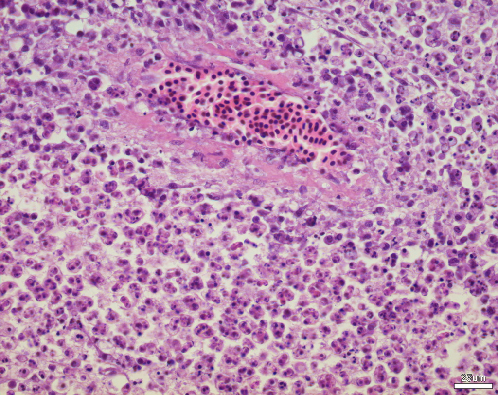
 | Slide # 58 Liver Mycobacteria H&E 200X.jpg | All Pathogens | |
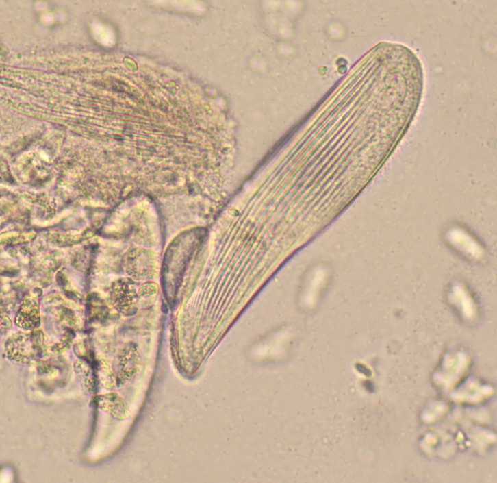
 | database_SLH0027 - Free ciliates in seawater x20.jpg | All Pathogens, Fact Sheet Images | |
 | DigsFish_29-nov-12-Pontobdella-loricata.jpg | All Pathogens, Contributed Images | shark - Carcharhinus sp. |
 | BO_Ichthyophonus_x20_38.jpg | All Pathogens | |
 | MYX_XX15_05a_x100bf_1shot.jpg | All Pathogens, Fact Sheet Images | |
 | Cshasta03.jpg | All Pathogens, Fact Sheet Images | |
 | Loma2.jpg | All Pathogens, Fact Sheet Images | |
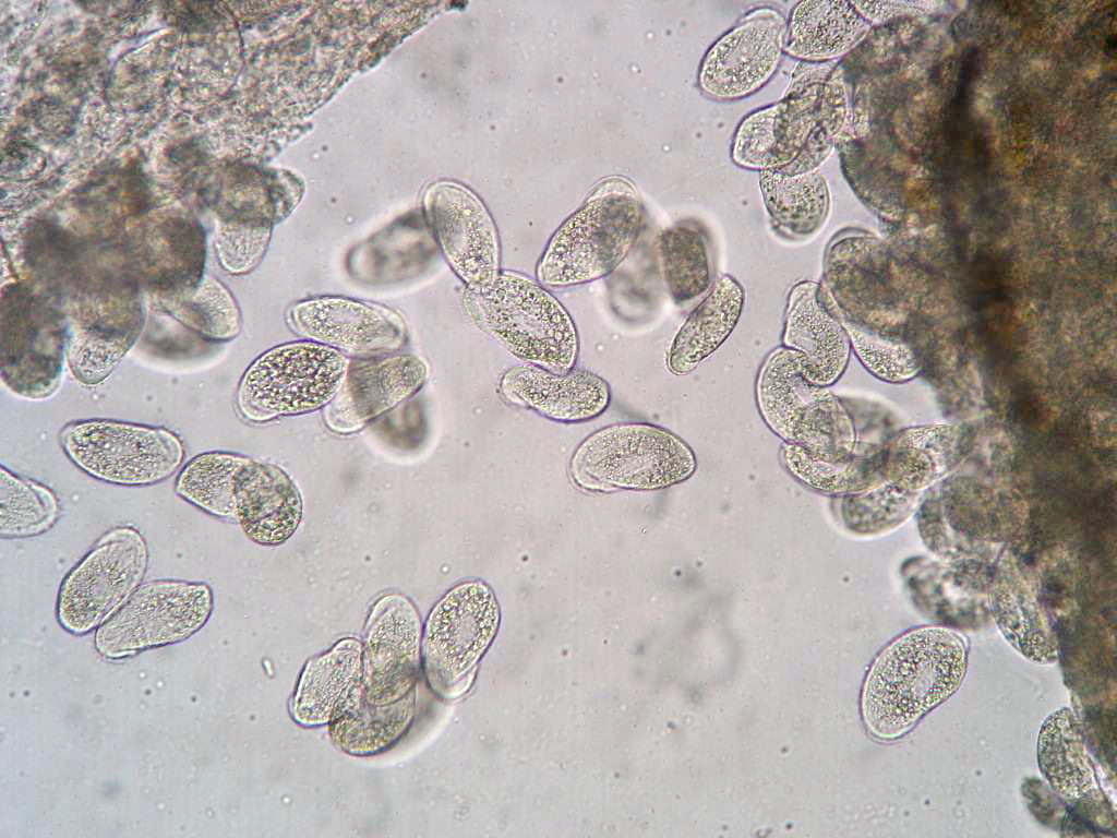
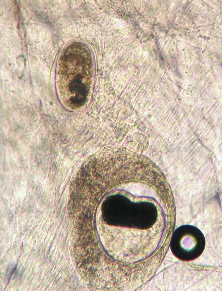
 | nanophyetus_salmonicola_CB08.jpg | All Pathogens, Fact Sheet Images | |