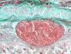
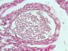
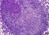
 | BO_Loma_x63_18.jpg | All Pathogens | |
 | MYX_XX15_05a_x100bf_1shot.jpg | All Pathogens, Fact Sheet Images | |
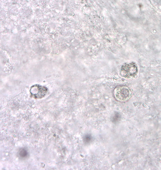
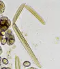
 | Cshasta03.jpg | All Pathogens, Fact Sheet Images | |
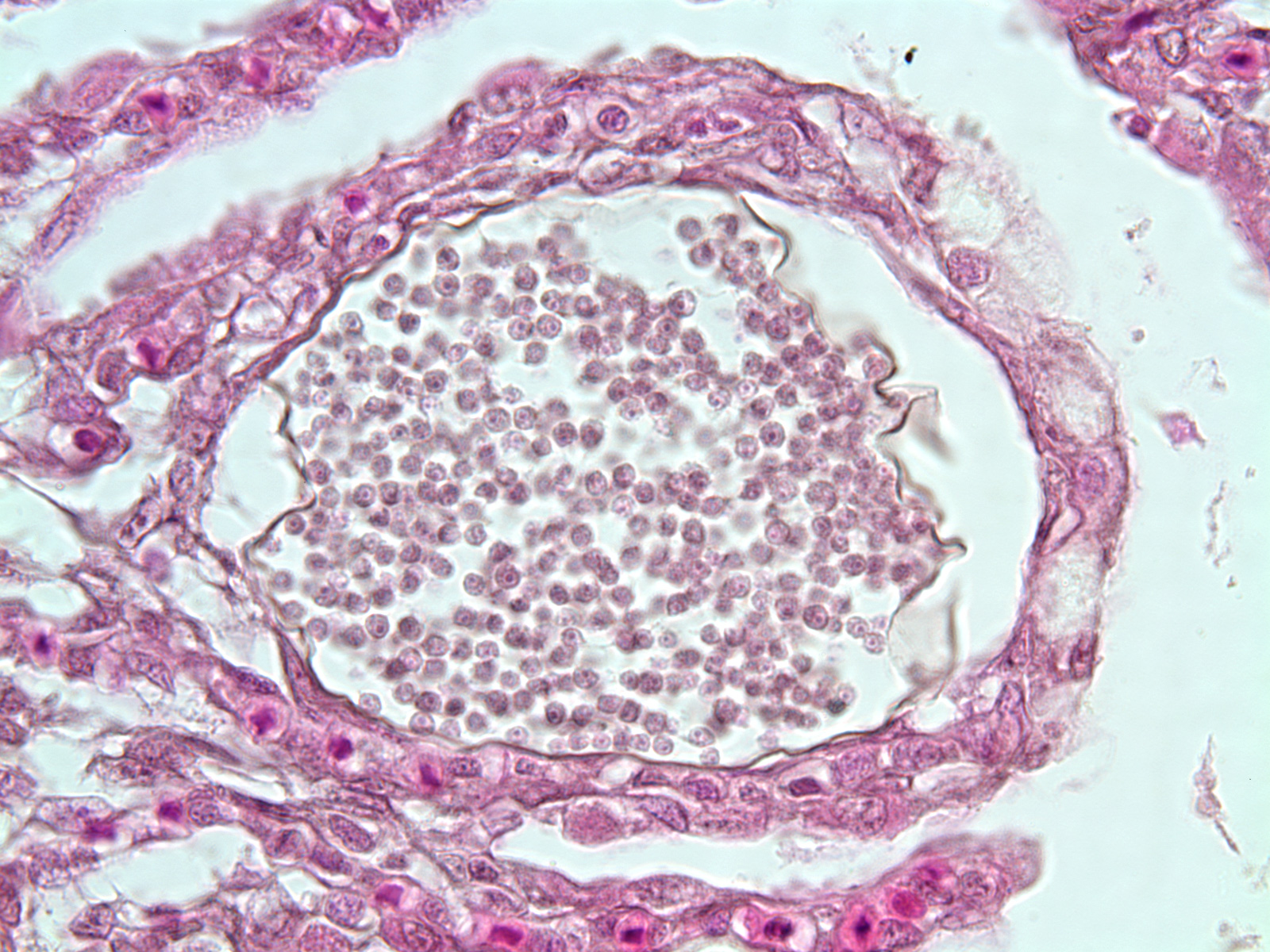
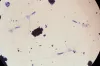
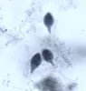
 | Loma2.jpg | All Pathogens, Fact Sheet Images | |
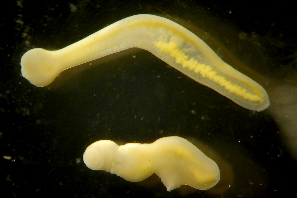
 | nanophyetus_salmonicola_CB08.jpg | All Pathogens, Fact Sheet Images | |
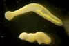
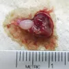 | P1020307.jpg | All Pathogens, Fact Sheet Images | bluegill |
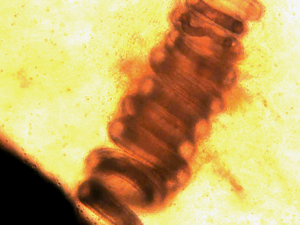
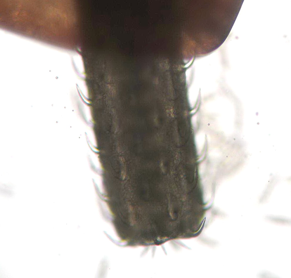
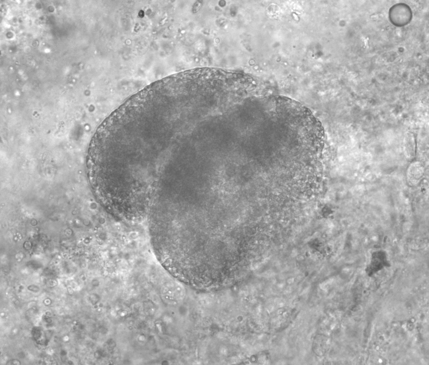
 | Glochidia_03.jpg | All Pathogens, Fact Sheet Images | |
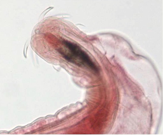
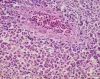
 | nematode_Contracecum_Bass_Nematode_CB01.jpg | All Pathogens, Fact Sheet Images | largemouth bass |
 | nematode_CB04.jpg | All Pathogens, Fact Sheet Images | |
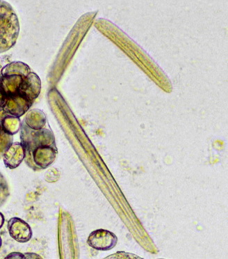
 | nematode_Salvelinema_walkeri_CB05.jpg | All Pathogens, Fact Sheet Images | |
 | Acanthocephala_CB04.jpg | All Pathogens, Fact Sheet Images | |
 | Acanthocephala_Echinorhynchus_lageniformis_CB04.jpg | All Pathogens, Fact Sheet Images | |
 | Acanthocephala_Neoechinorhynchus_sp_CB07.jpg | All Pathogens, Fact Sheet Images | |
 | trematode_Clinostomum_marginatum_CB04.jpg | All Pathogens, Fact Sheet Images | |
 | tricophyra_CB02.jpg | All Pathogens, Fact Sheet Images | |
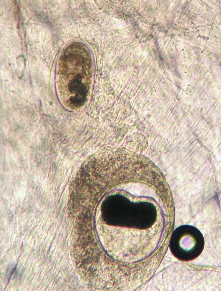
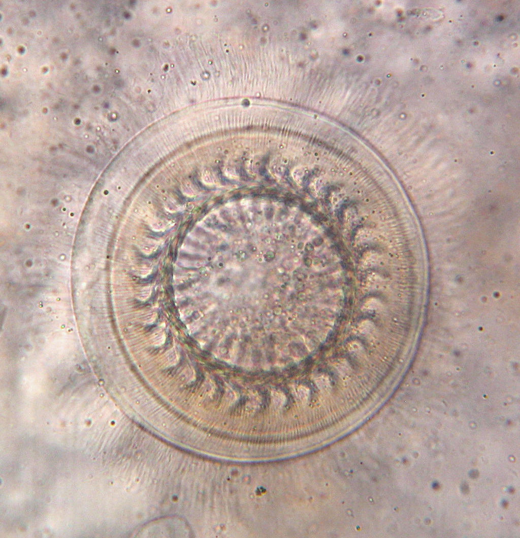
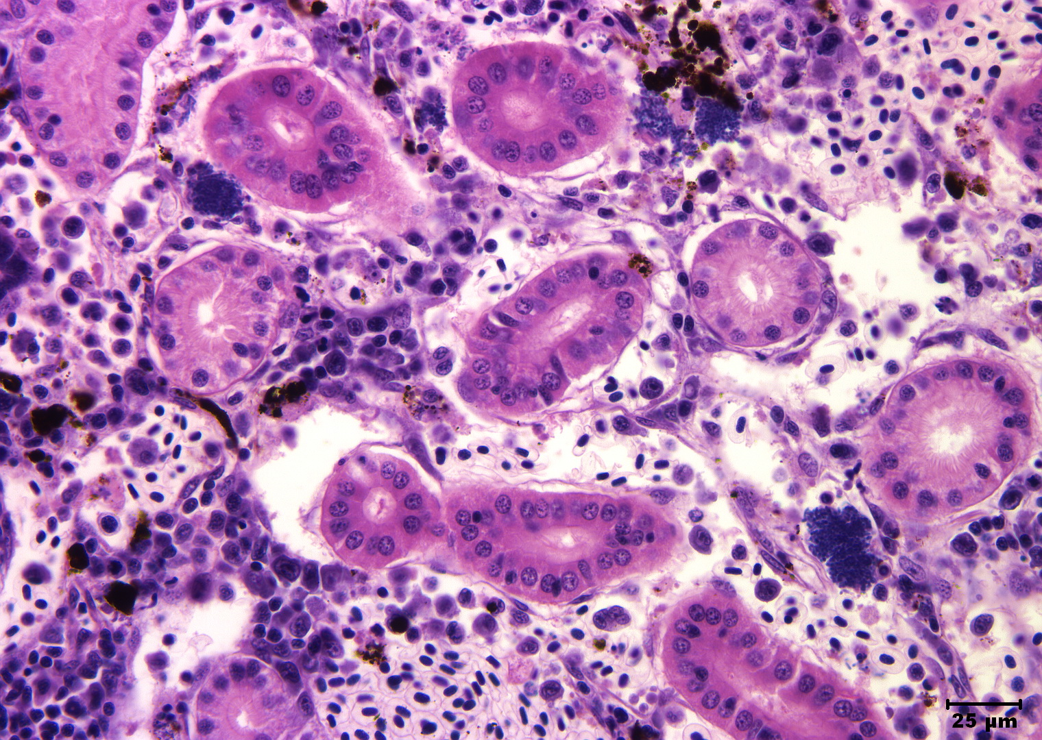
 | Minsidiosus_CB01.jpg | All Pathogens, Fact Sheet Images | |
 | SA_Loma_02b_x40bf_4shot.jpg | All Pathogens, Fact Sheet Images | |
 | CB_Trich7.jpg | All Pathogens, Fact Sheet Images | |
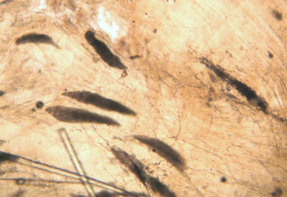
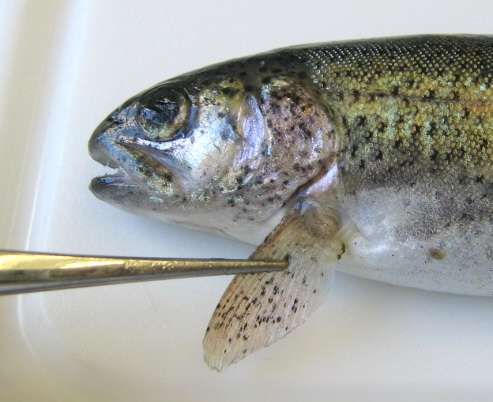
 | CB_BLk_SPOT2.jpg | All Pathogens, Fact Sheet Images | rainbow trout |
 | CB_Gill_amoeb33.jpg | All Pathogens, Fact Sheet Images | |
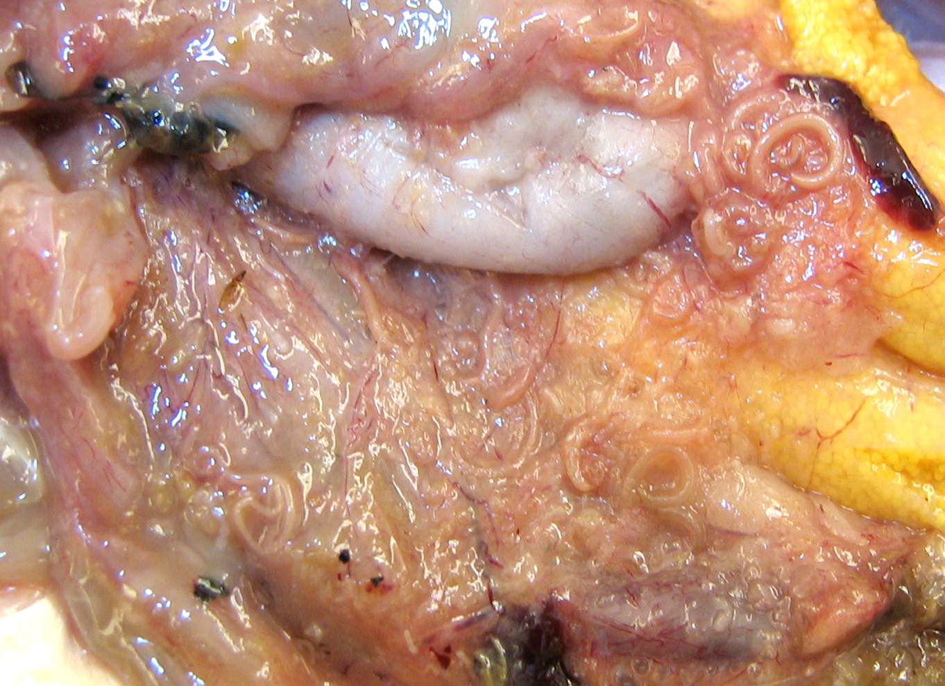
 | SLH_DSC00212.jpg | All Pathogens | Tubifex |
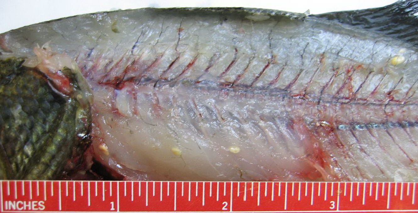
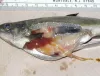
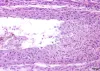
 | P1000375ed.jpg | All Pathogens, Fact Sheet Images | smallmouth bass |
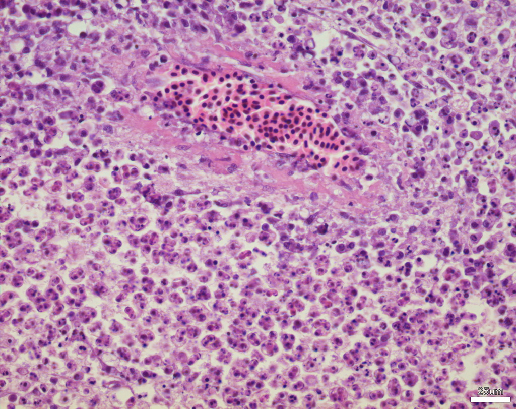
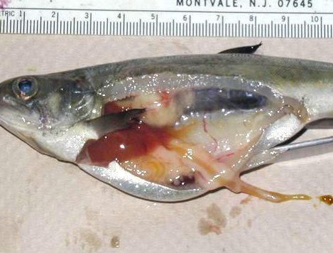
 | CB_BKD_02_ChS.jpg | All Pathogens, Fact Sheet Images | Spring Chinook salmon |
 | IHNV_kaufman1.jpg | All Pathogens, Fact Sheet Images | |
 | BO_Dermocystidium_x63_25.jpg | All Pathogens, Fact Sheet Images | |
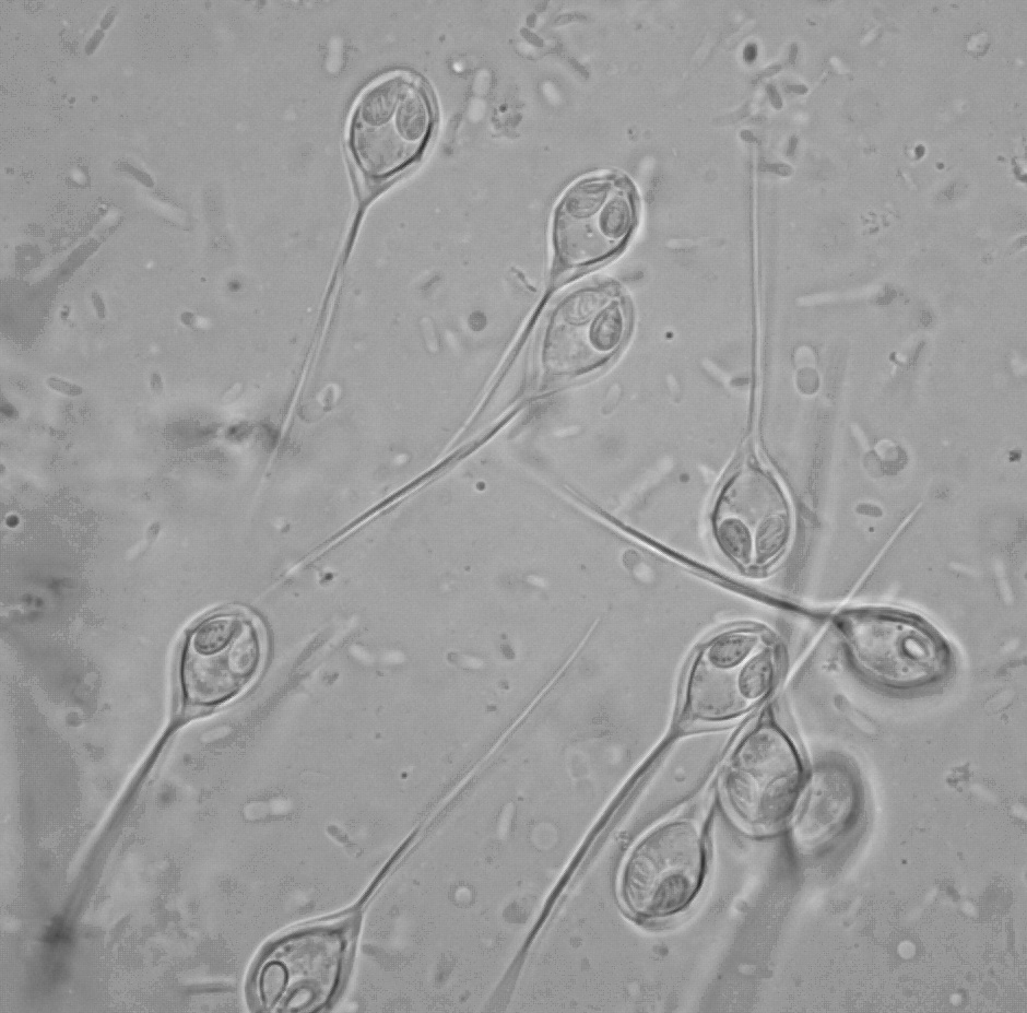
 | BO_Hexamita_x63_36.jpg | All Pathogens, Fact Sheet Images | |
 | Slide-#-100-Gills-Koi-Herpes-Virus-H&E-400X.jpg | All Pathogens | common carp |
 | Slide # 18 Kidney Aeromonas salmonicida H&E 400X.jpg | All Pathogens | |
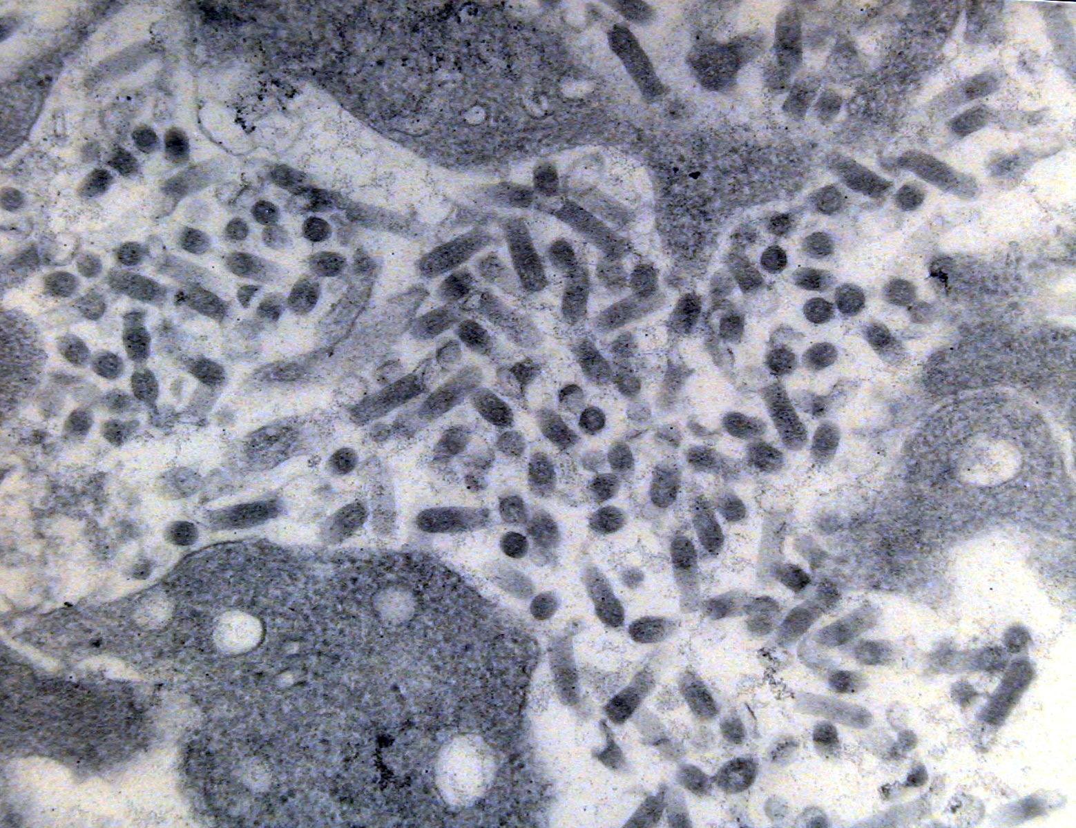
 | Slide # 58 Liver Mycobacteria H&E 400X.jpg | All Pathogens | |
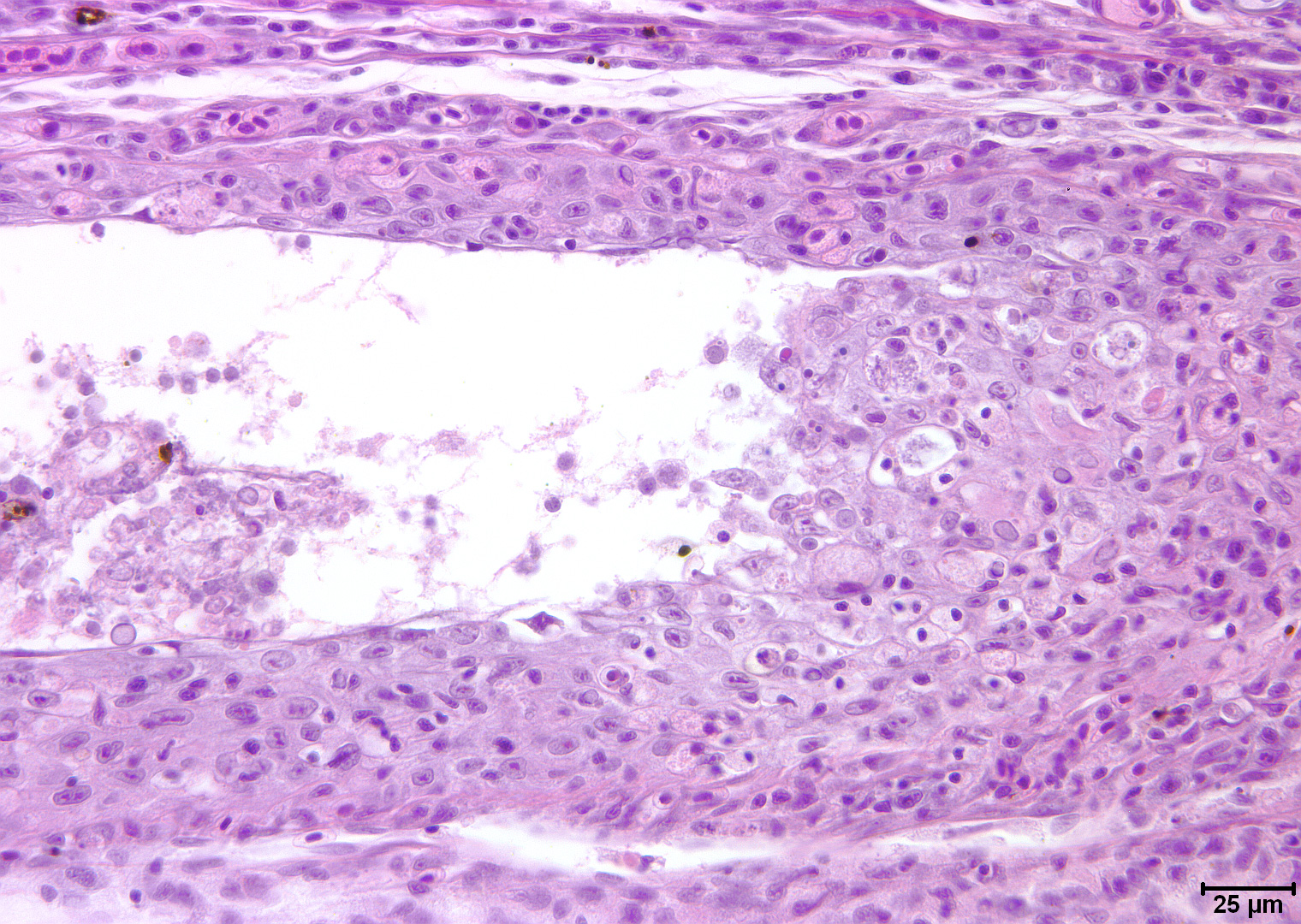
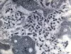
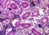
 | database_SLH0028 - Double infection.jpg | All Pathogens, Fact Sheet Images | |